Search results (66 results)
-
 Valsalva Retinopathy
Oct 28 2024 by Andrew Jin, MD
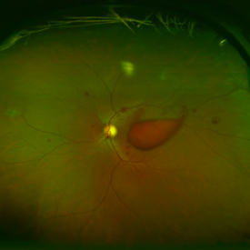
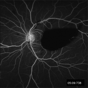
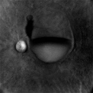
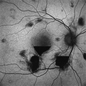
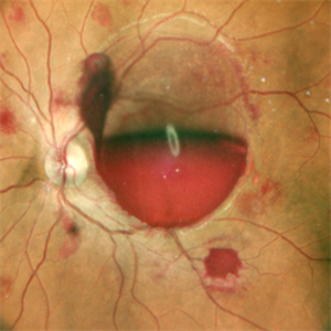
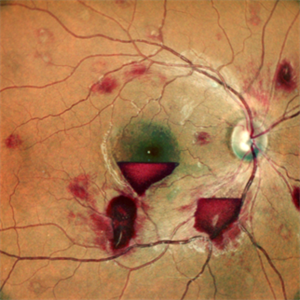
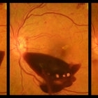
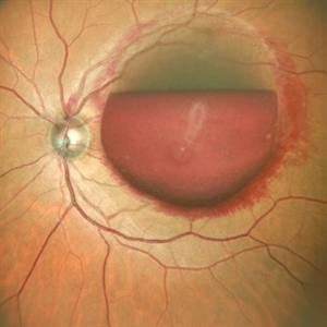
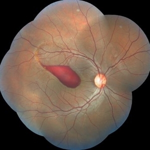
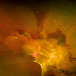
These photos depict the fundus photo and corresponding fluorescein angiogram for a 43 year old man with emesis after food poisoning. Note the blockage from the central preretinal hemorrhage and scattered peripheral intraretinal hemorrhages.
Condition/keywords: fluorescein angiogram (FA), fundus photograph, valsalva retinopathy
-
 Valsalva Retinopathy
Oct 28 2024 by Andrew Jin, MD
These photos depict the fundus photo and corresponding fluorescein angiogram for a 43 year old man with emesis after food poisoning. Note the blockage from the central preretinal hemorrhage and scattered peripheral intraretinal hemorrhages.
Condition/keywords: fluorescein angiogram (FA), fundus photograph, valsalva retinopathy
-
 Pre-Retinal Hemorrhage With Disc Edema
Apr 19 2024 by Akansha Sharma
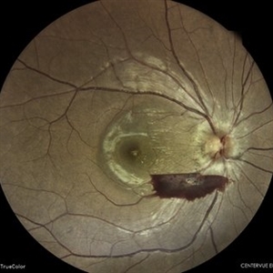
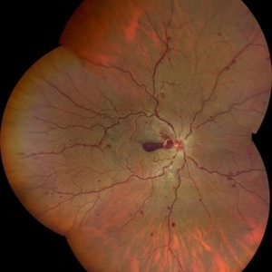
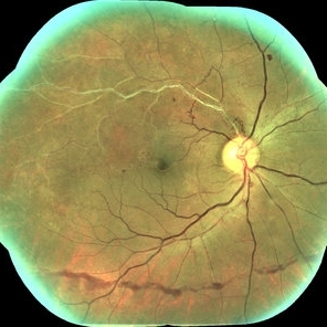
Color fundus photograph of a 39 year old female with disc edema along with a pre-retinal hemorrhage.
Photographer: Dr. Akansha Sharma, Bharati Eye Hospital
Condition/keywords: disc edema, preretinal hemorrhage
-
 Preretinal Hemorrhage
Sep 29 2023 by Angela Rico
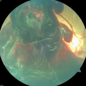
48 y/o male with Severe PDR
Photographer: Angela Rico M.D.
Condition/keywords: hemorrhage
-
 High risk Proliferative Diabetic Retinopathy treated with Pan Retinal Photocoagulation
Nov 5 2022 by Somnath Chakraborty, MD
A Fundus Photo Montage of 43 year old Asian Male with Type 2 Diabetes Mellitus since 7 years who presented with sudden onset diminition of vision in his Left eye. BCVA OS was 20/200. He was diagnosed to have Pre retinal bleed due to Proliferative Diabetic Retinopathy and was treated with Pan Retinal Photocoagulation. This image shows a large neo-cascular frond at the disc and superior to it with Pre-retinal bleed and Fresh laser marks along
Photographer: Pulak Roy
Condition/keywords: diabetic blindness, diabetic retinopathy vitrectomy study (DRVS), fresh laser burns, laser photocoagulation, preretinal hemorrhage, proliferative diabetic retinopathy (PDR)
-
 SUB-HYALOID HEMORRHAGE
Oct 20 2022 by Akansha Sharma
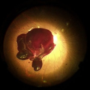
RETRO IMAGE OF A 23 YEAR OLD MALE WITH SUBHYALOID HEMORRHAGE IN A CASE OF ANAEMIC RETINOPATHY
Photographer: Dr. Akansha Sharma-Retina Foundation, Ahmedabad
Condition/keywords: anaemic retinopathy, preretinal hemorrhage, subhyaloid hemorrhage
-
 SUBHYALOID HEMORRHAGE
Oct 20 2022 by Akansha Sharma
AUTOFLUORESCENCE IMAGE OF A 23 YEAR OLD MALE WITH SUBHYALOID HEMORRHAGE IN A CASE OF ANAEMIC RETINOPATHY
Photographer: Dr. Akansha Sharma-Retina Foundation, Ahmedabad
Condition/keywords: anaemic retinopathy, preretinal hemorrhage, subhyaloid hemorrhage
-
 SUB HYALOID HEMORRHAGE
Oct 20 2022 by Akansha Sharma
COLOUR FUNDUS PHOTO OF A 23 YEAR OLD MALE WITH SUBHYALOID HEMORRHAGE IN A CASE OF ANAEMIC RETINOPATHY
Photographer: Dr. Akansha Sharma-Retina Foundation, Ahmedabad
Condition/keywords: anaemic retinopathy, preretinal hemorrhage, subhyaloid hemorrhage
-
 SUB HYALOID HEMORRHAGE
Oct 20 2022 by Akansha Sharma
COLOUR FUNDUS PHOTOGRAPH OF A 23 YEAR OLD MALE WITH SUB HYALOID HEMORRHAGE IN A CASE OF ANAEMIC RETINOPATHY
Photographer: Dr. Akansha Sharma-Retina Foundation, Ahmedabad
Condition/keywords: anaemic retinopathy, preretinal hemorrhage, subhyaloid hemorrhage
-
 YAG Laser in Preretinal Hemorrhage
Sep 27 2021 by Bruno André Cardoso Pereira
Fundus photograph series of a 55-year-old man with left eye preretinal hemorrhage involving the visual axis, resolved with NdYAG laser. Photographs taken before, 1 hour after and 1 month after the laser.
Photographer: Bruno Pereira
Imaging device: Topcon TRC-50DX
Condition/keywords: diabetic retinopathy, NdYAG laser, preretinal hemorrhage
-
 Macroaneurysm
Jun 23 2021 by Cláudia Farinha
Color Optomap from a middle-aged man with preretinal hemorrhage due to a macroaneurysm.
Photographer: Claudia Farinha, MD
Imaging device: Optomap, Optos
Condition/keywords: macroaneurysm, preretinal hemorrhage
-
 CRVO With Preretinal Hemorrhage
Mar 16 2021 by MOHIT GUPTA
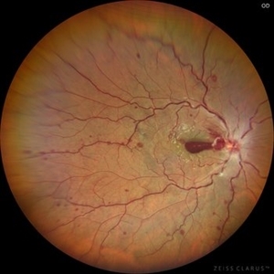
Fundus photograph of right eye of a young male after 2nd dose of Covishield vaccine presented to us with central retinal vein occlusion and preretinal hemorrhage at macula in right eye.
Photographer: Dr Mohit Gupta
Imaging device: Zeiss Clarius
Condition/keywords: central retinal vein occlusion (CRVO), preretinal hemorrhage
-
 Central Retinal Vein Occlusion with Preretinal Hemorrhage
Mar 16 2021 by MOHIT GUPTA
Fundus photograph of right eye of a young male after 2nd dose of Covishield vaccine presented to us with central retinal vein occlusion and preretinal hemorrhage at macula in right eye.
Photographer: Dr Mohit Gupta , Prakash Netra Kendr, Lucknow, India
Imaging device: zeiss clarus
Condition/keywords: central retinal vein occlusion (CRVO), preretinal hemorrhage
-
 Premacular Subhyaloid Hemorrhage
Jan 20 2021 by Nivesh Gupta
A 41-year-old male patient complaining of diminution of vision in left eye since 6 days. His best corrected visual acuity finger counting at 2 meters.
Photographer: Nivesh Gupta, Retina Fellow, Retina Foundation, Ahmedabad, India
Condition/keywords: hypertensive retinopathy, preretinal hemorrhage, subhyaloid hemorrhage
-
 Pre Macular Subhyaloid Hemorrhage
Jan 20 2021 by Nivesh Gupta
A 41 year old male patient complaining of diminution of vision in left eye since 6 days. His best corrected visual acuity finger counting at 2 meters.
Photographer: Nivesh Gupta, Retina Fellow, Retina Foundation, Ahmedabad, India
Condition/keywords: hypertensive retinopathy, preretinal hemorrhage, subhyaloid hemorrhage
-
 Valslava Retinopathy
Jan 15 2021 by Priya Rasipuram Chandrasekaran, MBBS, DO, DNB, FRCS
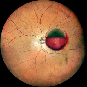
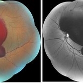
This is the fundus photo and red free montage showing preretinal hemorrhage of the left eye along the superior retina, abutting the disc margin and extending as far as the macula. There are few scattered flame shaped hemorrhages superiorly, nasally and inferiorly with a central white spot mimicking Roth spots.
Condition/keywords: valsalva retinopathy
-
 Mixed Occlusion of Artery and Vein
Jan 6 2021 by Renata Garcia Franco, Md
Male with a history of smoking, sudden low vision of the right eye, retinal neovascularization and inferior preretinal hemorrhage.
Photographer: Fatima Hernandez, Instituto de la Retina del Bajio SC
Imaging device: Zeiss
Condition/keywords: arterial occlusion
-
 OCT Showing Premacular Hemorrhage
Nov 26 2020 by Priya Rasipuram Chandrasekaran, MBBS, DO, DNB, FRCS
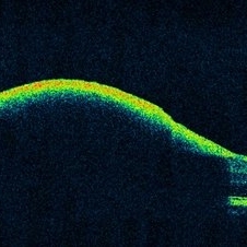
A 26-year-old male with no h/o trauma or underlying systemic disease presented with the complaint of central scotoma in the right eye since 1 month and fundus examination showed preretinal hemorrhage in the supero-temporal quadrant extending into the macular area and OCT macula showing premacular hemorrhage.
Condition/keywords: premacular hemorrhage
-
 Preretinal Hemorrhage Extending into the Macula
Nov 26 2020 by Priya Rasipuram Chandrasekaran, MBBS, DO, DNB, FRCS
A 26-year-old male with no h/o trauma or underlying systemic disease presented with the complaint of central scotoma in the right eye since 1 month and fundus examination showed preretinal hemorrhage in the supero-temporal quadrant extending into the macular area and OCT macula showing premacular hemorrhage.
Condition/keywords: preretinal hemorrhage
-
 Massive Commotio Retinae
Oct 20 2020 by Veronika Yehezkeli
Fundus photograph of a 24-year-old male, made after blunt trauma with a plastic bottle. Note massive commotio retinae and preretinal hemorrhages in the contralateral to trauma area.
Photographer: Veronika Yehezkeli, Meir medical center, Israel
Condition/keywords: blunt trauma, commotio retinae, preretinal hemorrhage, trauma
-
 Massive Commotio Retinae
Oct 20 2020 by Veronika Yehezkeli
Fundus photograph of a 24-year-old male, made after blunt trauma with a plastic bottle. Note massive commotio retinae and preretinal hemorrhages in the contralateral to trauma area.
Photographer: Veronika Yehezkeli, Meir medical center, Israel
Condition/keywords: blunt trauma, commotio retinae, preretinal hemorrhage
-
 Massive Commotio Retinae
Oct 20 2020 by Veronika Yehezkeli
24-year-old man was injured from an explosion of a plastic bottle towards the nasal conjunctiva of his left eye. A massive commotio retinae was diagnosed superotemporally.
Photographer: Veronika Yehezkeli, Meir medical center, Israel
Condition/keywords: blunt trauma, commotio retinae, preretinal hemorrhage
-
 Dense Preretinal Hemorrhage
May 15 2020 by Iuri Golubev, MD
34-year-old male w/h/o DM type 1 and PDR.
Condition/keywords: preretinal hemorrhage, proliferative diabetic retinopathy (PDR)
-
 Proliferative Sickle Cell Retinopathy
Apr 30 2020 by Jordan M Burnham, MD
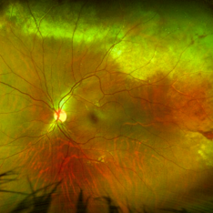
This ultra-widefield fundus photo of the right eye demonstrates proliferative sickle cell retinopathy resulting in severe visual loss for a young man eventually requiring vitrectomy. Central vitreous hemorrhage and subhyaloid hemorrhage covers the macula (white arrow), causing profound vision loss. A fibrotic, regressed, sea fan neovascularization complex is present in the temporal periphery (green arrow). Subretinal fluid is present in the temporal retinal periphery within the area between the fibrosed sea fan lesion and the posterior preretinal hemorrhage (yellow arrow), likely due to traction or a retinal break obscured by the heme.
Condition/keywords: sickle cell retinopathy
-
 Severe Proliferative Diabetic Retinopathy
Mar 2 2020 by Anfisa Ayalon, MD
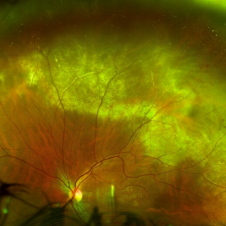
Fundus photograph of a 40-year-old patient with diabetes. Note severe fibrovascular membrane that covers almost the whole posterior pole, areas of tractional retinal detachment and preretinal hemorrhages.
Photographer: Anfisa Ayalon, MD., Meir Medical Center, Kfar Saba, Israel.
Imaging device: California, Optos 200 DTX
Condition/keywords: hemorrhage, neovascularization (NV), proliferative diabetic retinopathy (PDR), tractional retinal detachment
 Loading…