Search results (304 results)
-
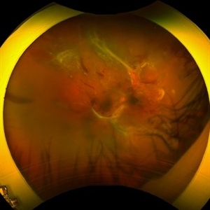
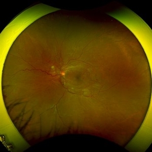
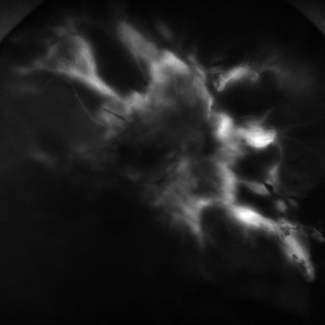
 All That Glows Yellow Isn’t Mellow: Coats' Disease Unveiled
Nov 4 2025 by SHRADDHA RAJ SHRIVASTAVA
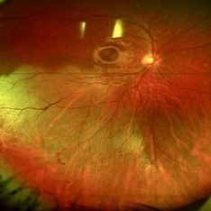
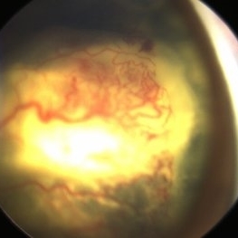
Montage fundus image of an 11 year old boy diagnosed with left eye Coats' disease (stage 3A1), reveals a hyperemic disc and surrounding intra-retinal exudates superior to the disc. There is a single fibroglial nodule at the macula causing submacular fibrosis with exudation. We can see areas of pigmentary changes and RPE atrophy in posterior pole and mid-peripheral retina supero-temporally. There is massive yellowish subretinal exudation in all the quadrants, which are associated with telangiectatic aneurysmal capillary dilation, more prominently seen in the nasal periphery. Supero-nasally we can also see an orange-red elevated vaso-proliferative mass with overlying dilated capillaries, which has likely developed secondary to untreated long standing disease. We can also see associated extrafoveal subtotal exudative retinal detachment in the inferior and nasal quadrants.
Photographer: Dr. Shraddha Raj Shrivastava
Imaging device: Nidek Mirante SLO/OCT (Confocal scanning/Spectral domain OCT)
Condition/keywords: COATS DISEASE, exudative detachment, leukocoria, subretinal exudates, Xanthocoria, yellow exudate
-
 Commotio Retinae
Aug 7 2025 by Gabriel Costa Andrade, PhD
Color fundus photograph of a 13-year-old girl who was hit by accidental discharge of gel bullet in her right eye. She presented with retinal whitening with intraretinal hemorrhages in temporal inferior area of the peripheral retina.
Photographer: Gabriel Andrade
Condition/keywords: macula, Retina, Trauma
-
 Combined Occlusion with TRD
Jun 30 2025 by Shivankar Sen, MS, FVRS
Posterior Pole and Ultra-wide field Fluorescein angiogram of a 79 yr. old one eyed male revealing arterial occlusion, grossly non-perfused peripheral retina with neovascularisation elsewhere and significant tractions at the posterior pole.
Photographer: Gayathri M S, Dr. Shivankar Sen MD
Imaging device: Heidelberg Spectralis HRA+OCT
Condition/keywords: arterial occlusion, Traction retinal detachment, Vein Occlusion
-
 Ocular Ischemic Syndrome
Jun 18 2025 by Korey Starkey
58-year-old patient with OIS in both eyes. Patient has had PRP in the past, however, presence of NVD with peripheral nonperfusion remains despite PRP.
Photographer: Korey Starkey
Imaging device: Optos
Condition/keywords: DME, FA early phase, fluorescein angiogram (FA), NVD, ocular ischemic syndrome, ois, Optos, peripheral retinal nonperfusion
-
 LCA type 12
Apr 10 2025 by Joshua Friedman
LCA type 12 due to pathogenic mutations in RDH12. 13-year-old male with a visual acuity of 20/80 and 20/300 in the right and left eye, respectively. There is extensive pigment migration in the peripheral retina and macula. Like RPE65, there is widespread hypoautofluorescent signal, however, the peripapillary retina is uniquely spared in this form of LCA. On OCT, there is almost complete loss of the retina centrally.
Photographer: Stephen Tsang, MD, PhD
Condition/keywords: Leber Congenital Amaurosis
-
 Advance Coats' Disease
Feb 15 2025 by Theinchai Pasurakul, MD
From the fundus image, the peripheral retina exhibits telangiectatic vessels accompanied by light bulb aneurysms at their terminal ends.
Photographer: Michael J. Shapiro MD, Advocate Lutheran General Hospital, Des Plaines
Imaging device: Retcam
Condition/keywords: Coats' disease, light-bulb aneurysms
-
 Familial Exudative Vitreoretinopathy
Jan 17 2025 by Aditya S Kelkar, MS, FRCS, FASRS,FRCOphth
A fundus photograph of a 16-year-old boy reveals temporal peripheral retinal non-perfusion and incomplete vascularization.
Photographer: Optom Mansi Raut
Imaging device: Optos Daytona
Condition/keywords: familial exudative vitreoretinopathy (FEVR)
-
 A Classic Case of Retinal Ora Serrata Imaging
Jan 16 2025 by yuan duo
A 5-year-old girl, born full-term with no history of systemic disease, presented with poor vision since early childhood and underwent fundus examination. Anterior segments of both eyes showed no significant abnormalities. Fundus examination revealed retinal folds extending from the optic disc to the temporal peripheral retina, with blood vessels coursing through the folds (A, B). Avascular zones were observed in the peripheral retina, and the ora serrata’s boundaries were clearly visible, displaying dentate processes and bays (C, D). Retinal pigmentation was evident. Genetic testing confirmed the final diagnosis of bilateral Familial Exudative Vitreoretinopathy (FEVR).
Condition/keywords: Retinal Ora Serrata
-
 Familial Exudative Vitreoretinopathy
Jan 16 2025 by yuan duo
A 5-year-old girl, born full-term with no history of systemic disease, presented with poor vision since early childhood and underwent fundus examination. Anterior segments of both eyes showed no significant abnormalities. Fundus examination revealed retinal folds extending from the optic disc to the temporal peripheral retina, with blood vessels coursing through the folds (A, B). Avascular zones were observed in the peripheral retina, and the ora serrata’s boundaries were clearly visible, displaying dentate processes and bays (C, D). Retinal pigmentation was evident. Genetic testing confirmed the final diagnosis of bilateral Familial Exudative Vitreoretinopathy (FEVR).
Condition/keywords: Retinal Ora Serrata
-
 Atypical Tubercular Occlusive Peripheral Retinal Vasculitis
Jun 21 2024 by Tejaswita Verma
Follow up right eye fundus photograph of a 27 year old male with vision 6/12 , diagnosed with systemic tuberculosis(mediastinal lymphadenopathy on chest CT) on oral steroids, and started on ATT .We can see a parafoveal sub-ILM hemorrhage with vascular sheathing and hemorrhages in inferior and temporal quadrants . The patient was advised anti-VEGF intravitreal injection, later sectoral laser after resolution of inflammation
Photographer: DR. TEJASWITA VERMA
Imaging device: MIRANTE
Condition/keywords: obliterative peripheral vasculitis, ocular tuberculosis
-
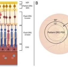 Summary of Retinal Origins of Various Electroretinographic Components
May 13 2024 by Gabrielle Hallai
Each layer of the retina can be assessed using various electroretinographic techniques. A) A summary of the retinal origins of each response is shown here. B) Macular function can be assessed by pattern and multifocal ERG recording and provide a specific assessment of central vision, while flash tests are dominated by the more peripheral retina.
Photographer: Gabrielle Hallai, PhD, Cleveland Clinic Cole Eye Institute
Condition/keywords: electroretinography, ERG
-
 Peripheral Retinal Degeneration (L-ORD)
Apr 17 2024 by Virginia Gebhart
92 year old female with bilateral patchy, sharply demarcated circular areas of chorioretinal atrophy with hyperpigmented margins in the mid to far periphery. Labs showed normal plasma ornithine levels ruling out generalized gyrate atrophy. Also intermediate uveitis and CMD/CME. FTA-ABS, Quant gold, and HLA-A29 labs all negative.
Photographer: Virginia Gebhart
Imaging device: Optos California
Condition/keywords: cystoid macular degeneration, cystoid macular edema (CME), FA, Fluorescein angiography, peripheral retinal degeneration
-
 Phoenix
Feb 21 2024 by Sayena . Jabbehdari, MD, MPH, MBA
A 60-year-old Caucasian female presented with reduced night vision and constricted visual fields. The fundus exam revealed pigmentary changes in the peripheral retina. Fundus autofluorescence depicted the schematic appearance of a Phoenix , with the hypo-autofluorescence corresponding to the head and wings of the phoenix. Genetic testing was positive for a heterozygous RHO mutation
Photographer: Sayena Jabbehdari MD MPH
Condition/keywords: retinitis pigmentosa
-
 Macular Hole-RRD
Feb 19 2024 by Ogugua Ndubuisi Okonkwo, MD, FRCS (Edin), FASRS
Fundus photograph of a 60-year-old male myope, who has multiple retina tears with rolled edges in the peripheral retina, associated with a macular hole and retinal detachment.
Photographer: Zainab Ogunsanu, Eye Foundation Hospital & Eye Foundation Retina Institute, Lagos.
Imaging device: ZEISS CLARUS 700
Condition/keywords: Retinal detachment with macular hole
-
 Peripheral retinal degenerations
Jan 29 2024 by Anupama Kiran Kumar
Fundus photo of a young man who underwent barrage laser after he presented to the clinic with floaters and was diagnosed to have lattices with horse shoe tears and retinal holes.
Photographer: Dr Anupama Kiran Kumar DNB FVR , Narayana Nethralaya Bangalore
Imaging device: Mirante SLO/OCT (Nidek Co., Gamagori, Japan)
Condition/keywords: lattice degeneration, peripheral retinal degeneration
-
 Severe Proliferative Diabetic Retinopathy
Jan 10 2024 by Ahmad B. Tarabishy, MD
33 year old female with 1 month history of vision loss right eye. Severe PDR was noted with VH and a TRD with severe FVP present OD.
Photographer: Sharon Story, Lakeland Eye Clinic
Imaging device: Optos
Condition/keywords: diabetic blindness, fibrovascular proliferation, nonperfusion diabetic retinopathy, peripheral retinal nonperfusion, proliferative diabetic retinopathy (PDR), tractional retinal detachment
-
 Alagille Syndrome UWF Color
Dec 4 2023 by Isaac Ezon, MD
43 yo Female with known Alagille Syndrome, referred for peripheral retinal changes. Subjective nyctalopia but no other symtpoms. Alagille Syndrome UWF Color.
Photographer: Tara Murray
Imaging device: Optos
Condition/keywords: hereditary choroidal dystrophy, hereditary retinal degeneration
 Loading…