Search results (80 results)
-
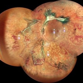
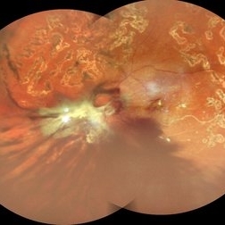 Table Top Tractional Retinal Detachment With Vitreous Hemorrhage in a Case of Proliferative Diabetic Retinopathy
Sep 12 2025 by Akansha Sharma
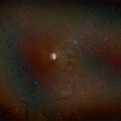
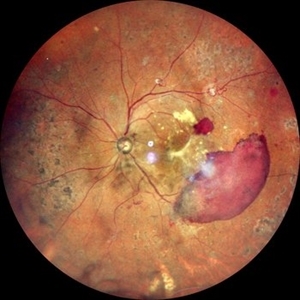
Color fundus photograph of a 56 year old male with table top tractional retinal detachment with vitreous hemorrhage in a case of proliferative diabetic retinopathy.
Photographer: DR. AKANSHA SHARMA
Condition/keywords: pan-retinal photocoagulation (PRP), PDR, proliferative diabetic retinopathy (PDR), PRP, TABLE TOP TRD, tractional retinal detachment, TRD, VH, vitreous hemorrhage
-
 Vasoproliferative Tumor (FEVR) s/p PPV/PRP
Aug 27 2025 by Virginia Gebhart
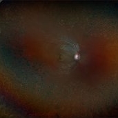
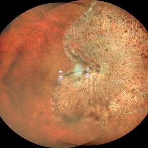
39 year old female with an amelanotic vascular lesion inferotemporal with CR atrophy inferior edge and likely lipid exudate superior edge. Pt presented with vitreous and sub-hyaloid hemorrhage. Findings from exam, ultrasound, FA all consistent with FEVR, stage 2. PPV with PRP performed, pt vison has improved from CF@2ft at initial visit to 20/100 PH 20/60 at 1 week post-op. Pt's 2 children have been recently examined with identical findings of FEVR
Photographer: Virginia Gebhart, Retina Consultants of Carolina
Imaging device: Optos California
Condition/keywords: familial exudative vitreoretinopathy (FEVR), pan-retinal photocoagulation (PRP), Vasoproliferative Tumor
-
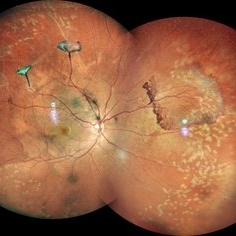
 Unstable PDR s/p Laser
Aug 4 2025 by Anjana Mirajkar, MS Ophthalmology
Fundus photograph of a 60 year old male with an unstable PDR showing traction at the posterior pole with sub hyaloid hemorrhage. Peripheral PRP marks can be seen.
Photographer: Dr. Anjana Mirajkar- HV Desai eye hospital ,Pune
Imaging device: Optos
Condition/keywords: pan-retinal photocoagulation (PRP), proliferative diabetic retinopathy (PDR), subhyaloid hemorrhage, tractional retinal detachment
-
 Proliferative Diabetic Retinopathy S/P Pan Retinal Photocoagulation
Mar 4 2025 by Prithvi Chandrakanth
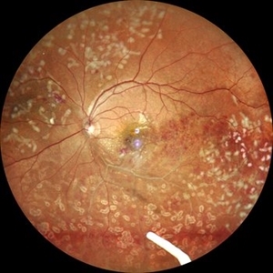
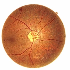
A 52-year-old female patient presented with complaints of diminishing vision, compounded by uncontrolled diabetes mellitus. Her Fundus examination revealed proliferative diabetic retinopathy, characterized by neovascularization of the disc and elsewhere, and sclerosed vessels. To address this, Pan Retinal Photocoagulation was performed, and the condition stabilized, halting the progression of the disease.
Photographer: DR PRITHVI CHANDRAKANTH, DR CHANDRAKANTH NETHRALAYA, KOZHIKODE, KERALA, INDIA
Imaging device: EIDON
Condition/keywords: Diabetic Retinopathy, Neovascularisation at the Disc (NVD), neovascularization of the disc (NVD), NVD, pan-retinal photocoagulation (PRP), PDR, PDR with NVE (periphery), PRP
-
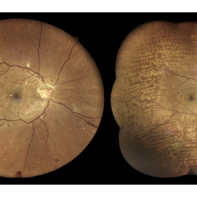
 Systemic Lupus Erythematosus (SLE) Vasculitis
Jan 29 2025 by Kimberly Wakester
Fundus photographs of an 13-year-old boy with Systemic Lupus Erythematosus (SLE) Vasculitis in both eyes s/p PRP laser. Patient is doing well s/p PRP Laser OU and with continued use of oral medications. Patient will be monitored with follow up exams to check for recurring vasculitis or recurring/worsening NVE/NVD. Patient is to continue ongoing management with Rheumatologist.
Photographer: Kimberly Wakester, COA
Imaging device: Optos California
Condition/keywords: NVD, NVE, occlusive vasculitis, pan-retinal photocoagulation (PRP), Systemic Lupus Erythematosus (SLE) Vasculitis
-
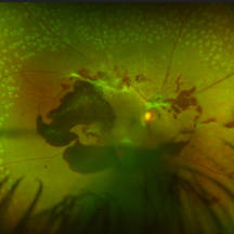
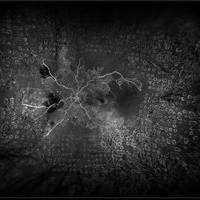
 Sickle-Cell Retinopathy
Jan 22 2025 by Virginia Gebhart
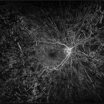
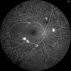
Fluorescein angiogram of 54 year old female with non-diabetic proliferative retinopathy. Recent labs confirm sickle-cell disease. FA shows temporal peripheral non perfusion with NV. S/p PRP with retrobulbar block
Photographer: Virginia Gebhart, Retina Consultants of Carolina
Imaging device: Optos California
Condition/keywords: FA, Neovascularisation elsewhere (NVE), non-perfusion, Nose, pan-retinal photocoagulation (PRP), PRP, sickle cell retinopathy
-
 FA- PRP
Dec 19 2024 by Angela Rico
68 y/o F with PRP
Photographer: Angela Rico M.D.
Condition/keywords: pan-retinal photocoagulation (PRP), PDR
-
 Idiopathic Retinal Vasculitis
Jun 9 2024 by Anjana Mirajkar, MS Ophthalmology
A widefield image of a 32 year old male of LE showing laser marks in inferior and superior half with an floating ozurdex implant (inferiorly) in a case of idiopathic retinal vasculitis.
Photographer: Dr. Anjana Mirajkar -Retina Foundation, Ahmedabad
Imaging device: Mirante-Nidek
Condition/keywords: idiopathic retinal vasculitis, laser photocoagulation, Ozurdex implant, pan-retinal photocoagulation (PRP)
-
 Proliferative diabetic retinopathy
Apr 28 2024 by Anjana Mirajkar, MS Ophthalmology
A widefield color image of a 60 year old male with type II diabetes showing sub hyaloid hemorrhage with traction at the fovea with hard exudates with venous looping along the supero temporal arcade with NVE inferiorly with surrounding laser marks.
Photographer: Dr. Anjana Mirajkar -Retina Foundation, Ahmedabad
Imaging device: Mirante-Nidek
Condition/keywords: pan-retinal photocoagulation (PRP), proliferative diabetic retinopathy (PDR)
-
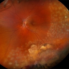
 Proliferative Diabetic Retinopathy
Apr 20 2024 by Tejaswita Verma
Widefield fundus photograph of the left eye of a 62 year old female with left eye lasered proliferative diabetic retinopathy showing neovascularisation elsewhere with few dot-blot hemorrhages.
Photographer: DR. TEJASWITA VERMA
Condition/keywords: Neovascularisation elsewhere (NVE), pan-retinal photocoagulation (PRP), proliferative diabetic retinopathy (PDR)
-
 Proliferative Diabetic Retinopathy
Apr 20 2024 by Tejaswita Verma
Fundus photograph of the left eye of a 62 year old female with lasered proliferative diabetic retinopathy showing neovascularisation elsewhere with few dot-blot haemorrhages
Photographer: DR. TEJASWITA VERMA
Condition/keywords: Neovascularisation elsewhere (NVE), pan-retinal photocoagulation (PRP), proliferative diabetic retinopathy (PDR)
-
 Serous Retinal Detachment in Advanced Proliferative Diabetic Retinopathy
Feb 15 2024 by Annaka Gooding
Ultra-Wide fundus photograph of a 29 year old female with a Serous Retinal Detachment in Advanced PDR. Patient present to clinic with LP vision following PPV and fill in PRP. Physician recommended oral prednisone treatment and to reassess at their following visit.
Photographer: Annaka Gooding, CPO
Imaging device: Optos California RGB
Condition/keywords: Diabetes, diabetic macular edema, fundus photography, OPTOS CALIFORNIA, pan-retinal photocoagulation (PRP), pars plana vitrectomy (PPV), proliferative diabetic retinopathy (PDR), serous retinal detachment, ultra-wide field imaging
-
 VITREOUS HEMORRHAGE WITH 360 DEGREE LASER PRP: RE
Jan 8 2024 by ANKIT JAIN
RIGHT EYE WIDEFILED FUNDUS PHOTO OF 38 YEARS OLD FEMALE WITH TYPE 1 DIABETES MELLITUS HAVING VITREOUS HEMORRHAGE WITH 360 DEGREE LASER PRP
Photographer: Dr Ankit Jain
Imaging device: MIRANTE
Condition/keywords: Diabetes, pan-retinal photocoagulation (PRP), PRP, vitreous hemorrhage
-
 LASER PRP: LE
Jan 8 2024 by ANKIT JAIN
LEFT EYE WIDEFILED FUNDUS PHOTO OF 38 YEARS OLD FEMALE WITH TYPE 1 DIABETES MELLITUS HAVING WITH 360 DEGREE LASER PRP
Photographer: DR ANKIT JAIN
Imaging device: MIRANTE
Condition/keywords: Diabetes, pan-retinal photocoagulation (PRP), PRP
-
 Ozurdex implant for diabetic macular oedema
Sep 6 2023 by PRATIK SHENOY, MBBS, DNB, FVRS
A 53-year-old female presented with diabetic macular oedema. She had undergone pan-retinal photocoagulation for proliferative diabetic retinopathy previously. She was injected with an intravitreal ozurdex implant for the same.
Photographer: Gaurav Kamble, Isha Netralaya
Imaging device: Optos
Condition/keywords: diabetic macular edema, Optos, ozurdex, pan-retinal photocoagulation (PRP), proliferative diabetic retinopathy (PDR)
-
 Regressed neovascularisation of disc
Jul 12 2023 by Prerana Shah
Neovascularisation of disc after Panretinal photocoagulation in proliferative diabetic retinopathy regressed or persistent
Photographer: Akshay Chavan , ShreeRamKrishna Netralaya, Thane ,India
Imaging device: Optopol Revo Fc
Condition/keywords: pan-retinal photocoagulation (PRP), proliferative diabetic retinopathy (PDR)
-
 Completed Pan-Retinal Fill-in Laser Photocoagulation in an Air filled eye at the end of Diabetic Vitrectomy Retina Surgery
Apr 28 2023 by Veer Singh, MS, FVRS, FMRF, FICO (Retina)
Completed Pan-Retinal Fill-in Laser Photocoagulation in an Air filled eye at the end of Diabetic Vitrectomy Retina Surgery
Photographer: Dr. Veer Singh
Condition/keywords: air-filled, pan-retinal photocoagulation (PRP), vitrectomy
-
 Intra-operative PRP in a diabetic vitrectomy case
Apr 28 2023 by Veer Singh, MS, FVRS, FMRF, FICO (Retina)
Pan-Retinal Fill-in Laser Photocoagulation in a Diabetic Vitrectomy Retina Surgery
Photographer: Dr. Veer Singh
Condition/keywords: diabetic, pan-retinal photocoagulation (PRP), vitrectomy
-
 Active diabetic retinopathy despite PRP
Oct 30 2022 by Diego Andrés Rodriguez, MD
A 52-year-old patient with active proliferative diabetic retinopathy despite good glycemic control and PRP performed 1 year ago in the right eye
Photographer: Sociedad de Cirugía Ocular
Imaging device: Clarus 700
Condition/keywords: diabetic retinopathy, pan-retinal photocoagulation (PRP), proliferative diabetic retinopathy (PDR), wide angle imaging
-
 STATUS POST PAN-RETINAL PHOTOCOAGULATION
Oct 11 2022 by Akansha Sharma
COLOUR FUNDUS PHOTO OF A 71 YEAR OLD MALE WITH SCARRING POST PAN-RETINAL PHOTOCOAGULATION
Photographer: Dr. Akansha Sharma-Retina Foundation, Ahmedabad
Condition/keywords: laser photocoagulation, pan-retinal photocoagulation (PRP)
-
 TRACTIONAL RETINAL DETACHMENT IN A CASE OF VASCULITIS
Mar 14 2022 by Akansha Sharma
MONTAGE OF A 27 YEAR OLD MALE WITH TREACTIONAL RETINAL DETACHMENT IN A CASE OF VASCULITIS
Photographer: Dr. Akansha Sharma-Retina Foundation, Ahmedabad
Condition/keywords: pan-retinal photocoagulation (PRP), tractional retinal detachment, VASCULITIS
-
 Central Retinal Vein Occlusion with Retinal Neovascularization
Jan 19 2022 by Olivia Rainey
Ultra-widefield fluorescein angiogram of a 56-year-old male with a Central Retinal Vein Occlusion with Retinal Neovascularization affecting his left eye. The patient presented on 1/19/2022 with scNLP vision in the left eye. The patient has good PRP, however areas of ischemia still remain untreated by laser. He also has severe neovascular glaucoma contributing to his poor vision.
Photographer: Olivia Rainey, OCT-C, COA
Imaging device: Optos California
Condition/keywords: central retinal vein occlusion (CRVO), FA early phase, hemorrhage, ischemic CRVO, left eye, neovascular glaucoma, Optos, pan-retinal photocoagulation (PRP), retinal ischemia, retinal neovascularization, ultra-wide field imaging
-
 Proliferative Diabetic Retinopathy after PRP Laser Treatment
Oct 3 2021 by Jesus Lozano, MD
Wide-field FAF Optos image of a 55-year-old male patient with high risk proliferative diabetic retinopathy presented with blurred vision in the right eye. Panretinal photocoagulation (PRP) treatment was performed in 2 sessions. Remanent epiretinal hemorrhage along the inferior arcade.
Photographer: Yair Bet Yosef, Hadassah Medical Center. Israel
Imaging device: Optos
Condition/keywords: laser photocoagulation, pan-retinal photocoagulation (PRP), proliferative diabetic retinopathy (PDR)
-
 Neovascularization in a Case of Idiopathic Vasculitis
Aug 18 2021 by ASRS Staff
Right eye, Wide field photograph Motage of 37 year-old male, having idiopathic vasculitis status post PRP. Patient's systemic evaluation was done, and everything was within normal limits.
Imaging device: Nidek Mirante
Condition/keywords: Idiopathic vasculitis, montage, neovascularization (NV), pan-retinal photocoagulation (PRP)
 Loading…