Search results (50 results)
-
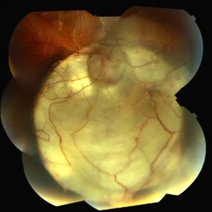
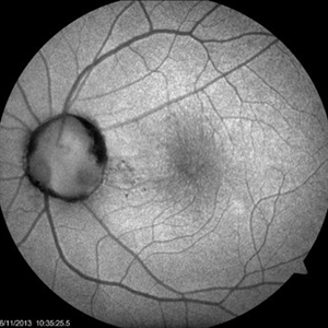
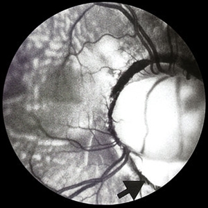
 Optic Nerve Coloboma and Rhegmatogenous Retinal Detachment
Sep 30 2025 by Píndaro Alonso Cruz-Benitez
Optic nerve coloboma and rhegmatogenous retinal detachment
Photographer: Píndaro Alonso Cruz-Benitez, APEC, Mexico
Condition/keywords: optic nerve coloboma, Retinal Detachment
-
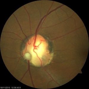
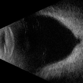 Posterior Staphyloma + ON-Coloboma
Aug 20 2025 by Gustavo Uriel Fonseca Aguirre
This axial B-scan reveals a highly myopic eye with a posterior staphyloma and an associated optic nerve coloboma. The staphyloma appears as a deep scleral outpouching adjacent to the optic disc, while the coloboma demonstrates a focal posterior excavation with retrobulbar extension.
Photographer: Gustavo U. Fonseca Aguirre, Hospital Conde de Valenciana, Ciudad de México
Condition/keywords: optic nerve coloboma, posterior staphyloma
-
 Optic Nerve Coloboma
Aug 14 2021 by Narciso F. Atienza, MD, MBA, FASRS, FPCS, FPAO.
19 year old male patient seen on routine examination for refraction. Had blurring of vision on the right eye since childhood. Was initially seen by a general ophthalmologist who diagnosed the patient with glaucoma. Present vision is CF at 3 feet uncorrected, and 20/400 with a refraction of -8.00 -1.50 X 180.
Photographer: Narciso F Atienza, Jr. MD MBA, FASRS, FPCS, FPAO. Legazpi Eye Center
Imaging device: Topcon TRC
Condition/keywords: optic nerve coloboma
-
 Chorioretinal Coloboma with Retinal Detachment
Dec 5 2020 by Niloofar Piri, MD
14-year-old female with 1q21.1 microdeletion syndrome and behavioral, intellectual, and systemic abnormalities, including congenital microcornea, iris coloboma, and chorioretinal and optic nerve coloboma presented with decreased vision. Right eye fundus taken with RetCam shows coloboma with retinal detachment. (Left eye showed white cataract with funnel RD on B-scan).
Photographer: Niloofar Piri MD, Douglas Snyder MD
Condition/keywords: chorioretinal coloboma, optic nerve coloboma
-
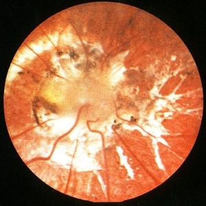
 Cat Eye Syndrome
Feb 11 2020 by Sophia El Hamichi, MD
A 3-year-old female with cat eye syndrome including iris, chorioretinal and optic nerve colobomas. Note the CNV temporally to the optic nerve coloboma (blue arrows)
Photographer: Giselle De Oliveira, Bascom Palmer Eye Institute, Miami
Imaging device: RetCam
Condition/keywords: cat eye syndrome, chorioretinal coloboma, choroidal neovascularization (CNV), coloboma, coloboma of optic disc, optic nerve coloboma
-
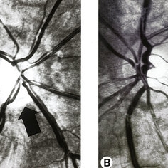
 Optic Nerve Coloboma Associated with Persistent Fetal Vasculature and Microphthalmia in the Setting of CHARGE Syndrome
Feb 3 2020 by Sophia El Hamichi, MD
An 8-month-old male referred for ophthalmic evaluation in the setting of CHARGE syndrome. EUA revealed microphthalmia with persistent fetal vasculature and optic disc coloboma OS (depicted in image A: fundus photograph and image B: fluorescein angiogram). OD exam revealed dysplastic microphthalmia.
Photographer: Abby Orcutt-Hayes, Murray Ocular Oncology and Retina
Imaging device: RetCam
Condition/keywords: CHARGE syndrome, microphthalmos, optic nerve coloboma, persistent fetal vasculature (PFV)
-
 Coloboma
Sep 7 2018 by John S. King, MD
11-year-old white female with bilateral optic nerve and retinochoroidal colobomas and an optic nerve pit in the right eye looking almost like pseudoduplication of the optic nerve. She is currently 20/30 OD and 20/20 OS. She has a history of laser by Dr. Zocchi about 10 years ago for a low lying, macula involving, serous retinal detachment, and has responded well.
Photographer: Stacey Coleman
Imaging device: Topcon
Condition/keywords: chorioretinal coloboma, inferior optic nerve coloboma, optic disc pit
-
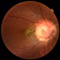
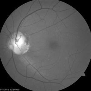
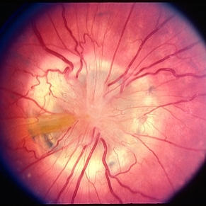
 Optic Nerve Coloboma
Nov 21 2014 by Thomas A. Ciulla, MD, MBA, FASRS
This 18-year-old woman has an optic nerve coloboma right eye with longstanding poor vision.
Photographer: Thomas Steele
Condition/keywords: coloboma of optic disc, coloboma of the optic nerve
-
 Retinal - Macular Coloboma
Mar 13 2014 by James B. Soque, CRA, OCT-C, COA, FOPS
Large retinal and optic nerve coloboma of a 31-year-old white male with 20/100 vision OS.
Photographer: James B Soque, CRA, COA
Imaging device: Topcon TRC 50DX, OIS 5 MP Camera,MERGE Software
Condition/keywords: coloboma of optic disc, color photo, macular coloboma
-
 Optic Nerve Coloboma an 2 Pits, Nasal and Temporal Blue Light
Nov 21 2013 by Alexandre Durao Alves Pereira, MD
Fundus photograph, color, red free, blue lite and FAF of a optic nerve coloboma with 2 pits, one nasal and other temporal.
Photographer: Alexandre Pereira
Imaging device: Visucam 300
Condition/keywords: optic nerve coloboma
-
 Optic Nerve Coloboma With 2 Pits, Nasal and Temporal Red Free
Nov 21 2013 by Alexandre Durao Alves Pereira, MD
Fundus photograph, color, red free, blue lite and FAF of a optic nerve coloboma with 2 pits, one nasal and other temporal.
Photographer: Alexandre Pereira
Imaging device: Visucam 300
Condition/keywords: optic nerve coloboma, red-free
-
 Optic Nerve Coloboma With 2 Pits, Nasal and Temporal Color
Nov 21 2013 by Alexandre Durao Alves Pereira, MD
Fundus photograph, color, red free, blue lite and FAF of a optic nerve coloboma with 2 pits, one nasal and other temporal.
Photographer: Alexandre Pereira
Imaging device: Visucam 300
Condition/keywords: color photo, optic nerve coloboma
-
 Optic Nerve Coloboma with 2 Pits, Nasal and Temporal FAF
Nov 21 2013 by Alexandre Durao Alves Pereira, MD
Fundus photograph, color, red free, blue lite and FAF of an optic nerve coloboma with 2 Pits, one nasal and other temporal
Photographer: Alexandre Pereira
Imaging device: Visucam 300
Condition/keywords: fundus autofluorescence (FAF), optic nerve coloboma
-
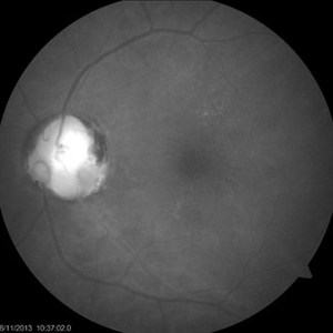
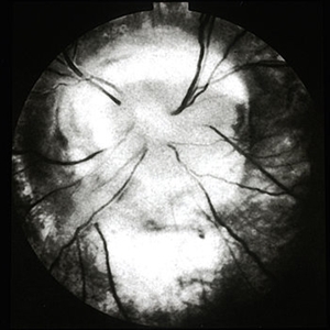
 Optic Nerve Coloboma
Feb 20 2013 by From the Collections of Thomas M. Aaberg, MD and Thomas M. Aaberg Jr., MD
No history; FA.
Condition/keywords: optic nerve coloboma
-
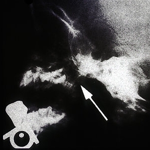
 Coloboma of Optic Nerve With Non-Rhegmatogenous Retinal Detachment
Feb 20 2013 by From the Collections of Thomas M. Aaberg, MD and Thomas M. Aaberg Jr., MD
21-year-old.
Condition/keywords: optic nerve coloboma
-
 ON coloboma
Feb 20 2013 by From the Collections of Thomas M. Aaberg, MD and Thomas M. Aaberg Jr., MD
No history or color photo.
Condition/keywords: black and white photo, optic nerve coloboma
-
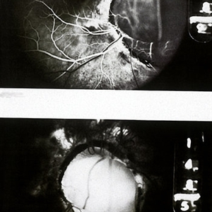
 Optic Nerve Coloboma / Morning Glory Syndrome / Encephalocele / X-ray
Feb 20 2013 by From the Collections of Thomas M. Aaberg, MD and Thomas M. Aaberg Jr., MD
Morning glory with associated encephalocele protruding into nasopharynx.
Condition/keywords: encephalocele, Morning Glory Syndrome, optic nerve coloboma, x-ray
-
 Morning Glory Optic Nerve; Coloboma
Feb 20 2013 by From the Collections of Thomas M. Aaberg, MD and Thomas M. Aaberg Jr., MD
No history.
Condition/keywords: Morning Glory Syndrome, optic nerve coloboma
-
 Morning Glory Syndrome
Feb 20 2013 by From the Collections of Thomas M. Aaberg, MD and Thomas M. Aaberg Jr., MD
18-month-old.
Condition/keywords: Morning Glory Syndrome, optic nerve coloboma
-
 Optic Nerve Coloboma / Morning Glory Syndrome / Encephalocele
Feb 20 2013 by From the Collections of Thomas M. Aaberg, MD and Thomas M. Aaberg Jr., MD
Morning glory with associated encephalocele protruding into nasopharynx.
Condition/keywords: encephalocele, Morning Glory Syndrome, optic nerve coloboma
-
 Morning Glory Syndrome – Dilitation and Contraction of Coloboma
Feb 20 2013 by From the Collections of Thomas M. Aaberg, MD and Thomas M. Aaberg Jr., MD
1) dilatation 2) contraction
Condition/keywords: Morning Glory Syndrome, optic nerve coloboma
 Loading…