Search results (625 results)
-
 Angiographic Storm: Fluorescein Leakage in Retinal Vasculitis
Nov 17 2025 by SHRADDHA RAJ SHRIVASTAVA
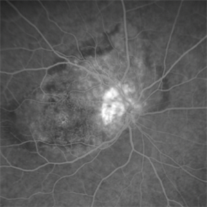
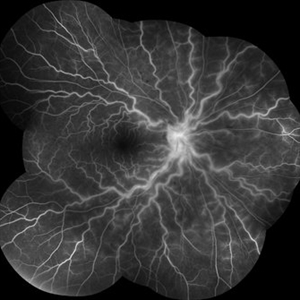
This left eye montage fundus fluorescein angiography (FFA) image of a 19 year old male with idiopathic retinal vasculitis, having skip vasculitic lesions predominantly involving retinal veins. There are areas of blocked fluorescence due to intraretinal hemorrhages, the involved veins have filling defects and occlusions, leading to formation of numerous collateral channels. The inflamed vessels also show perivascular fuzzy hyperfluorescent stain due to leakage of dye. We can also see multiple peripheral capillary non perfusion (CNP) areas, with a 'hot disc', suggestive of ongoing inflammation.
Photographer: Dr. Shraddha Raj Shrivastava
Imaging device: Nidek Mirante SLO/OCT (Confocal scanning/Spectral domain OCT)
Condition/keywords: FA late phase leakage, Fundus Fluorescein Angiography, idiopathic retinal vasculitis, optic disc leakage, VASCULITIS
-
 Neovascular Medusa: A Bad Hair Day at the Optic Disc
Nov 4 2025 by SHRADDHA RAJ SHRIVASTAVA
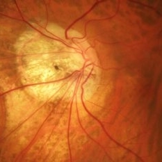
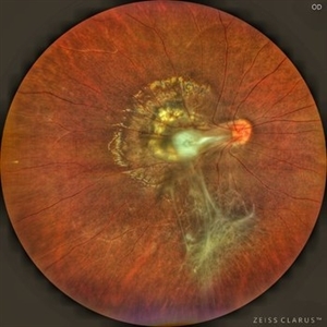
Left eye pseudocolor fundus photo of 67 year old male, diagnosed with both eyes proliferative diabetic retinopathy, showing hair-like fronds of active neovascularisation at the disc (NVD) extending into the vitreous, giving the medusa-head appearance. There is a band of fibrovascular proliferation nasal to the disc, with presence of hard exudates and dot hemorrhages at the macula.
Photographer: Dr. Shraddha Raj Shrivastava
Imaging device: Nidek Mirante SLO/OCT (Confocal scanning/Spectral domain OCT
Condition/keywords: Diabetic Retinopathy, fibrovascular proliferation, Neovascularisation at the Disc (NVD), proliferative diabetic retinopathy (PDR)
-
 Optic Disc Drusen in Rod Cone Dystrophy
Nov 3 2025 by Malvika Singh
Fundus autofluorescence of a 22 year old male with rod cone dystrophy with hyperautofluorescent disc drusen.
Photographer: Dr Malvika Singh, Retina Foundation, Ahmedabad, India
Imaging device: Mirante SLO/OCT
Condition/keywords: optic disc drusen, Rod cone dystrophy
-
 Peripapillary Neovessels
Oct 27 2025 by Oftalmontt Clínica Láser
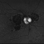
OCT-A of a 54-year-old male patient with Proliferative Diabetic Retinopathy, observed the large number of neovessels in the optic disc along with fibrovascular proliferation.
Photographer: Ophthalmic Medical Technologist
Imaging device: Avanti XR AngioVue OptoVue
Condition/keywords: diabetic retinopathy
-
 Hidden Papilla
Oct 16 2025 by Oftalmontt Clínica Láser
Tilted optic disc in a myopic patient.
Photographer: Ophthalmic Medical Technologist
Imaging device: Canon cx-1
Condition/keywords: myopia
-
 Juxtapapillary Retinal Capillary Hemangioblastoma
Oct 16 2025 by Sara Mayoral Sánchez
A hyperfluorescent lesion located superotemporal to the optic disc, consistent with a juxtapapillary retinal capillary hemangioma.
Photographer: Sara Mayoral Sánchez, H.U.Puerta del Mar, Cádiz
Condition/keywords: angiography with fluorescein, hemangioma, optic disc, retina capillary hemangioblastoma
-
 Retained PFCL Over the Optic Disc
Oct 14 2025 by rohan jain
Retained PFCL bubble over the optic disc after retinal detachment surgery.
Photographer: Dr. ROHAN JAIN
Imaging device: mirante
Condition/keywords: near infrared autofluorescence (NIRAF), PFCL
-
 Optic Nerve Drusen
Oct 8 2025 by Gustavo Uriel Fonseca Aguirre
This longitudinal B-scan demonstrates an optic nerve head drusen, appearing as a hyperechoic, well-defined focus at the optic disc with mild acoustic shadowing. The drusen exhibits a rounded contour and is superficial to the lamina cribrosa.
Photographer: Gustavo U. Fonseca Aguirre, Hospital Conde de Valenciana, Ciudad de México
Condition/keywords: optic nerve drusen
-
 Chorioretinal Coloboma
Oct 6 2025 by Seif Allah Anwar
Fundus photograph of the patient left eye showing large, well-demarcated, excavated chorioretinal coloboma involving the inferior fundus, extending from the optic disc to the periphery. The lesion appears white due to bare sclera visibility, with absence of overlying choroid and retina. Retinal vessels course over the colobomatous area inferiorly.
Photographer: Dr. Seif Anwar, FRCSEd
Imaging device: Centervue Eidon
Condition/keywords: chorioretinal coloboma
-
 Dropped Nucleus With Disc Pallor With Posterior Pole Retinal Detachment
Sep 12 2025 by Akansha Sharma
Color fundus photograph of a 60 year old male with a dropped nucleus with disc pallor with posterior pole retinal detachment.
Photographer: DR. AKANSHA SHARMA
Condition/keywords: dropped nucleus, fragmatome, nucleus drop, optic disc pallor, PALE DISC, POSTERIOR POLE RETINAL DETACHMENT, RD, retinal detachment
-
 Optic Disc Neovascularization
Sep 9 2025 by Seif Allah Anwar
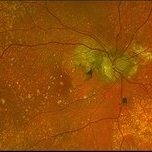
A case of high risk proliferative diabetic retinopathy with large disc neovascularization.
Photographer: Dr Seif Anwar
Imaging device: Topcon
Condition/keywords: optic disc neovascularization
-
 Large Leaking Optic Disc Neovascularization
Sep 9 2025 by Seif Allah Anwar
Large leaking optic disc neovascularization.
Photographer: Dr Seif Anwar
Imaging device: Topcon
Condition/keywords: Fundus Fluorescein Angiography
-
 Unexpected Sanctuary: Gas Bubble Entrapment in Morning Glory Disc
Sep 5 2025 by Danny Salgado Gómez
Fundus photograph of a 62-year-old male patient with Morning Glory syndrome in the right eye, who underwent vitrectomy, gas, and endolaser for posterior pole detachment. In the postoperative period, a gas bubble is observed within the optic disc, which persisted even after complete reabsorption of the intraocular gas.
Photographer: Dr. Danny Salgado, Retina and Vitreous Fellow, Clínica Oftalmológica del Caribe, Colombia.
Condition/keywords: gas bubble, intraocular gas, Morning Glory, Retinal Detachment, vitrectomy
-
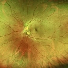
 Fluorescein Angiography Papillophlebitis Salauno
Sep 3 2025 by Pablo Angel Garcia Uribe
In the arteriovenous phase, fluorescein angiography demonstrated venous engorgement and tortuosity, with relative incompetence of the venous walls leading to mild leakage. Optic disc staining with late leakage was also observed. There was no evidence of significant capillary non-perfusion, and only subtle perivenous leakage was noted. The foveal region remained spared.
Photographer: Optom. Marilyn Alvarez Monroy, Clínica Oftalmológica Salauno
Imaging device: Visucam 524, Carl Zeiss Meditec AG, Jena, Germany
Condition/keywords: FA late phase leakage, retina
-
 The Retinal Tempest: Toxocara's Trail
Aug 31 2025 by Giriraj Vibhute
In this striking image, a central white granuloma spirals outward from the optic disc, surrounded by fibrous traction bands and scarring—the telltale markings of intraocular toxocara lesion. The retina is ravaged with proliferative vitreoretinal membranes and peripheral pigmentary changes, starkly illustrating the chronic inflammation and vision-threatening complications caused by Toxocara canis.
Photographer: Dr Giriraj Vibhute, MM Joshi eye institute, Hubli, India.
Condition/keywords: dragged disc, fibrous proliferation, Toxocara, toxocara canis
-
 Optic Disc Drusen
Aug 20 2025 by Drew Mitchell
Fundus Autofluorescence photo of an 86 year old woman with neovascular AMD with active CNV and optic disc drusen.
Photographer: Drew Mitchell OCT-C
Imaging device: Optos California
Condition/keywords: fundus autofluorescence (FAF), neovascular age-related macular degeneration (AMD), optic disc drusen, OPTOS
-
 Optic Disc Drusen
Aug 20 2025 by Drew Mitchell
Optos color photo of a 86 year old woman with neovascular AMD with active CNV and optic disc drusen.
Photographer: Drew Mitchell OCT-C
Imaging device: Optos California
Condition/keywords: color photo, optic disc drusen, OPTOS
-
 Optic Disc Drusen
Aug 20 2025 by Drew Mitchell
HD 1 line 100x scan through optic disc drusen. ODD are defined as Hyporeflective structures with a full or partial hyperreflective margin.
Photographer: Drew Mitchell OCT-C
Imaging device: Zeiss Cirrus 6000
Condition/keywords: OCT, optic disc drusen
-
 Posterior Staphyloma + ON-Coloboma
Aug 20 2025 by Gustavo Uriel Fonseca Aguirre
This axial B-scan reveals a highly myopic eye with a posterior staphyloma and an associated optic nerve coloboma. The staphyloma appears as a deep scleral outpouching adjacent to the optic disc, while the coloboma demonstrates a focal posterior excavation with retrobulbar extension.
Photographer: Gustavo U. Fonseca Aguirre, Hospital Conde de Valenciana, Ciudad de México
Condition/keywords: optic nerve coloboma, posterior staphyloma
-
 Persistent Hyperplastic Primary Vitreous Associated With Retrolental Fibrovascular Membrane
Aug 8 2025 by Pablo Angel Garcia Uribe
A 12-year-old Mexican male, asymptomatic, referred for evaluation after incidental finding of partial leukocoria on routine ophthalmologic examination. Slit-lamp evaluation revealed a fibrovascular retrolental membrane without evidence of retinal traction, associated with a fibrous stalk connecting to the optic disc. The stalk showed near-complete involution, consistent with a remnant of persistent fetal vasculature (posterior type).
Photographer: Pablo Angel García-Uribe, Clínica Oftalmológica Salauno, Mexico City
Condition/keywords: Persistent Hyperplastic Primary Vitreous Fibrovascular membrane
-
 Optic Disc Melanocytoma Hyperpigmented Magnocellular Nevus of the Optic Disk (HMNOD)
Aug 5 2025 by SHRADDHA ASHOK CHANDORKAR, DNB DO FVRS
OCT DISC image of a case of optic nerve melanocytoma.
Photographer: Dr.Shraddha A Chandorkar
Imaging device: zeiss
Condition/keywords: optic disc melanocytoma
-
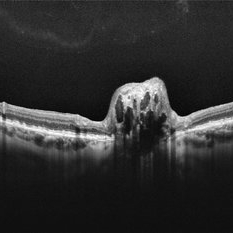
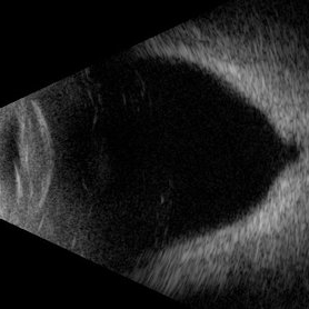
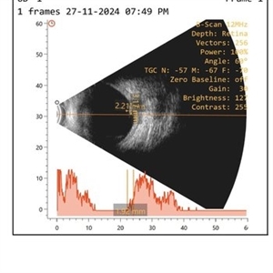 Optic Disc Melanocytoma USG Measurements
Aug 5 2025 by SHRADDHA ASHOK CHANDORKAR, DNB DO FVRS
B Scan showing measurements of mass in a case of optic nerve melanocytoma.
Photographer: Dr.Shraddha A Chandorkar
Condition/keywords: b scan
-
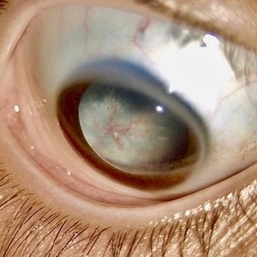
 Black Swan - Optic Disc Melanocytoma
Aug 5 2025 by SHRADDHA ASHOK CHANDORKAR, DNB DO FVRS
Just like the Black Swan which signifies an event that comes as a surprise, can have a major effect, and is often inappropriately rationalized after the fact with the benefit of hindsight, a 50 yr old presbyopic lady came to OPD with complains of diminution of vision - BCVA being 6/6 N6 in both eyes. Fundus examination revealed a pigmented nodule covering the optic disc .In most cases, fluorescein angiography of a melanocytoma of the optic disk demonstrates hypofluorescence throughout the angiogram. OCT disc showed elevated lesion, OCT macula normal and USG B scan with measurements were done to corroborate the posterior extension and to note increase in size if any on follow ups, perimetry was done to check for any field defects. All tests seemingly within normal limits - Patient was counselled and asked for 6 monthly follow up. Optic Disc Melanocytoma usually unilateral known to be a benign lesion that carries an excellent prognosis, the malignancy of this specific condition is rare 1-2%. The mean age at diagnosis of optic disk melanocytoma is 50 years with a median of 52 and range of 1–91 years. It is possible that melanocytoma is a congenital lesion but may not become clinically apparent until later in life, perhaps due to acquisition of pigment in a previously amelanotic lesion.
Photographer: Dr.Shraddha A. Chandorkar
Imaging device: topcon
Condition/keywords: optic disc melanocytoma
-
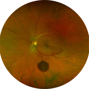 Large Subhyaloid Hemorrhage
Jul 11 2025 by Jessilla Phou
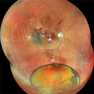
This is a fundus photograph depicting a large subhyaloid hemorrhage in the mid periphery of the left eye. The patient, a 53-year-old female, presented with a sudden onset of floaters, headache, and blurred vision. The image also demonstrates associated optic disc hemorrhage, vitreous hemorrhage, retinal hemorrhage, and venous tortuosity. Despite the extensive workup performed and the severity of the hemorrhage, no underlying cause was determined.
Photographer: Jessilla Phou
Imaging device: Optos California
Condition/keywords: fundus photograph, optic disc hemorrhage, retinal hemorrhage, venous tortuosity, vitreous hemorrhage
-
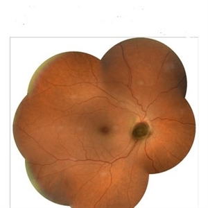
 Pseudoduplication of the Optic Disc
Jul 9 2025 by Hrishikesh Naik, MS
A peripapillary colobomatous pseudo-duplication of the optic disc as seen in an asymptomatic 23 year old female with myopia referred for routine retinal periphery screening. Rest retinal exam was normal. Duplication of the optic disc can be classified as either true duplication or pseudoduplication, both of which are rare clinical conditions. Pseudodoubling of the optic disc is commonly caused by optic disc or peripapillary colobomas, characterized by a circumscribed, disc-like lesion with radiating vessels but only one normal optic nerve. A few cases have involved pathological myopia, moderate myopia, proliferative diabetic retinopathy and CHARGE syndrome. The lesion is often found inferior to the normal optic disc. The patient was advised regular follow ups.
Photographer: Hrishikesh Naik
Imaging device: Optos Daytona
Condition/keywords: Coloboma, Pseudoduplication of optic disc
 Loading…