Search results (6 results)
-
 Central Retinal Vein Occlusion with Macular Edema
Jan 29 2025 by Kimberly Wakester
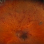
Fundus photograph of a 62-year-old man with central retinal vein occlusion with macular edema and a new PVD with an operculated retinal tear in the left eye. Laser to retinal tear was completed. Patient will return in 2-3 weeks for follow up exam with possible intravitreal injection for the CRVO with edema and to follow up on the operculated retinal tear s/p retinal tear laser.
Photographer: Kimberly Wakester, COA
Imaging device: Optos California
Condition/keywords: central retinal vein occlusion (CRVO), operculated tear, PVD
-
 Operculated Hole with Barrier Laser
Nov 5 2019 by Nichole Lewis
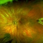
66-year-old female with an operculated retinal hole s/p barrier laser treatment. Choroidal Nevus Inferior.
Photographer: Nichole Lewis
Imaging device: Optos
Condition/keywords: barrier laser, choroidal nevus, operculated retinal hole, operculated tear
-
 Operculated Retinal Tear
Apr 8 2019 by Gary R. Cook, MD, FACS
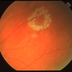
Fresh laser photocoagulation spots around an operculated retinal tear.
Imaging device: Topcon VT-50
Condition/keywords: laser photocoagulation, laser retinopexy, operculated tear, retinal tear
-
 Operculated Retinal Tear
Apr 8 2019 by Gary R. Cook, MD, FACS
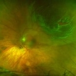
Fresh, operculated retinal tear with surface hemorrhage.
Imaging device: Topcon VT-50
Condition/keywords: hemorrhage, operculated tear, retinal tear
-
 Retinal Detachment With a Chronic Appearing Operculated Tear
Jun 21 2018 by Nichole Lewis
73-year-old male with a macula-on retinal detachment with a chronic appearing operculated tear. VA 20/30.
Photographer: Nichole Lewis
Condition/keywords: operculated tear
-
 Operculated Tear at the Back Surface
Dec 10 2012 by Yale L. Fisher, MD
In this ultrasound movie there is little movement of the vitreous. Observe the operculated, small, relatively strongly reflective tissue on the detached, mildly reflective, vitreous back surface (yellow arrow). There is a sagital view of the superior aspect of the lateral rectus muscle (orange arrow). Operculated tear at 10 o'clock position.
Condition/keywords: operculated tear, ultrasound, video, vitreous
 Loading…