Search results (52 results)
-
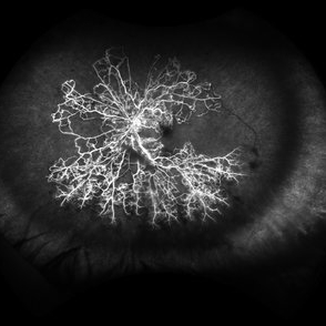
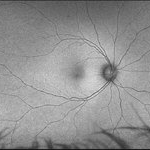
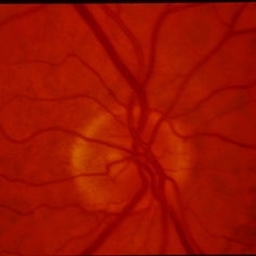
 Ocular Ischemic Syndrome
Jun 18 2025 by Korey Starkey
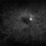
58-year-old patient with OIS and Hollenhorst plaque.
Photographer: Korey Starkey
Imaging device: Optos
Condition/keywords: capillary nonperfusion, fluorescein angiogram (FA), hollenhorst plaque, NVD, ocular ischemic syndrome, Optos
-
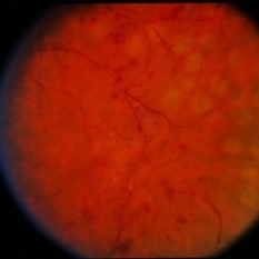
 Ocular Ischemic Syndrome
Jun 18 2025 by Korey Starkey
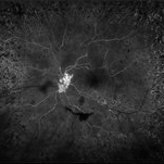
58-year-old patient with OIS in both eyes. Patient has had PRP in the past, however, presence of NVD with peripheral nonperfusion remains despite PRP.
Photographer: Korey Starkey
Imaging device: Optos
Condition/keywords: DME, FA early phase, fluorescein angiogram (FA), NVD, ocular ischemic syndrome, ois, Optos, peripheral retinal nonperfusion
-
 Treatment of Ocular Ischemic Syndrome with Hyperbaric Oxygen Therapy
Dec 2 2024 by Catherine S Kang
A 66-year-old female with past medical history significant for hypertension and ocular ischemic syndrome. She presented in emergency department (ED) reporting eye pain and blurred vision in both eyes since earlier that morning. On examination, best corrected visual acuity in each eye was counting fingers (20cm). Further investigation was performed and fluorescein angiography revealed a delay in choroidal filling. The patient has been followed for ocular ischemic syndrome since the onset of the condition and hyperbaric oxygen therapy was promptly initiated. Final best corrected visual acuity was 20/150 and macula developed atrophy.
Photographer: Catherine Kang
Condition/keywords: hyperbaric oxygen therapy, ocular ischemic syndrome
-
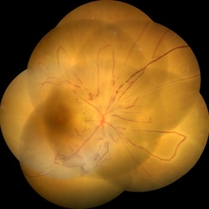
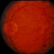
 Ocular Ischemic Syndrome
Jun 27 2023 by Mauricio Bayram-Suverza, MD
Fundus photograph of a 72-year-old man referred to the retina department due to long-term, painless vision loss in his left eye. Following fluorescein angiography, prolonged arteriovenous transit time and hypoperfusion were detected. Consequently, the patient was advised to undergo cardiological evaluation.
Photographer: Mauricio Bayram-Suverza, Fundación Hospital Nuestra Señora de la Luz
Imaging device: Optos California
Condition/keywords: Fluorescein angiography, ocular ischemic syndrome, Ultra-wide field retinal imaging
-
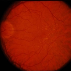
 Ocular Ischemic Syndrome/ Severe NPDR
Oct 6 2021 by Becca Harris
53 year old female with Severe NPDR and Ocular Ischemic Syndrome.
Photographer: Becca Harris
Imaging device: Optos California
Condition/keywords: Diabetic Retinopathy, left eye, nonproliferative diabetic retinopathy, ocular ischemic syndrome, optos, retinal ischemia
-
 Diabetic Retinopathy
Aug 15 2021 by JEFFERSON R SOUSA, Tecg.º (Biomedical Systems Technology)
A 32-year-old female patient attended the Suel Abujamra Institute (ISA) for evaluation and management. In the process of evaluating the visual picture, important retinal alterations, typically of diabetic retinopathy, were observed. As shown in the OCT-A (Angioplex) exam, extensive area of ischemic vascular incompentence.
Photographer: JEFFERSON R SOUSA - Centro de Estudos e Pesquisas Oftalmológicas Dr. Andre MV Gomes, Instituto Dr. Suel Abujamra São Paulo-Brasil
Imaging device: Angioplex protocol, OCT CIRRUS 5000. Zeiss
Condition/keywords: diabetic mellitus, diabetic retinopathy, ischaemic diabetic maculopathy, ischemia, ocular ischemic syndrome, retinopathy
-
 Ocular Ischemic Syndrome - Takayasus Disease
Jun 14 2021 by Raja Rami P Reddy, MD FRCS FASRS
16-year-old girl, a known case of Takayasus disease with history of stroke presented with unilateral cataract. The photograph is taken 2 days following cataract surgery. The fellow eye has normal fundus.
Photographer: Raja Rami Reddy P
Condition/keywords: ocular ischemic syndrome, Takayasus disease
-
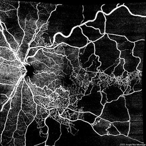
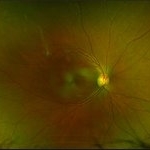
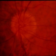
 Ocular Ischemic Syndrome
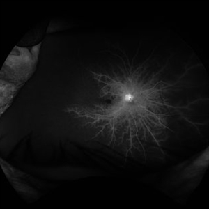
Aug 21 2020 by Gabriel Costa Andrade, PhD
Wide field fluorescein angiography of a 76-year-old male with ocular ischemic syndrome associated with severe stenosis of the left internal carotid artery.
Photographer: Gabriel Andrade
Imaging device: OPTOS - CALIFORNIA
Condition/keywords: ocular ischemic syndrome
-
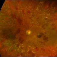
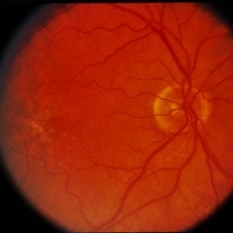
 Ocular Ischemic Syndrome
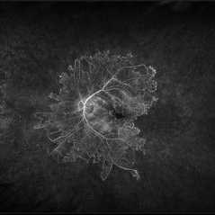
Jun 20 2018 by Andreas Ebneter, MD, PhD, FASRS
Ocular ischemic syndrome can present with a wide variety of ocular findings in both the anterior and posterior segments. The color fundus image of this 77-year-old male shows scattered blot hemorrhages in the deep retinal layers of the posterior pole that are only occasionally confluent. Commonly, these typical hemorrhages are predominantly found in the mid-periphery. Fluorescein angiography helps in confirming the diagnosis. Choroidal filling time is frequently somewhat delayed and patchy. Arteriovenous transit time is clearly prolonged. Staining of both veins and arteries in late images (top right) reflects diffuse endothelial cell damage with compromise of the blood-retina barrier. The peripheral retina is affected by extensive non-perfusion.
Photographer: Eva Steffen, Bern University Hospital, Switzerland
Imaging device: Optos 200Tx and Heidelberg Spectralis OCT
Condition/keywords: ocular ischemic syndrome
-
 Tractional vs Combined Tractional/Rhegmatogenous Retinal Detachment with Active Neovascularization OS
Jun 1 2018 by Hosam Attia, MD
47-year-old African American, with history of diabetes mellitus of unknown duration and control, was referred for initial evaluation for conjunctival laceration in his left eye, following accidental finger nail injury, 6 days prior to presentation. - On exam, his vision was 20/50 OD and Bare HM/ LP OS. - Fundus color photos OD: No significant pathology, aside from attenuated vasculature OS: Chronic, Mac-Off, almost closed funnel tractional vs combined tractional/rhegmatogenous retinal detachment with large neovascularization (NVE) superiorly, detached ghost vessels, mild fresh vitreous hemorrhage, sub-retinal bands and inferior white vitreous debris from old hemorrhage (not shown) - FA OD: No significant pathology aside from possible mild capillary non-perfusion in the extreme periphery, attenuated vasculature and possible tiny microaneurysms, nasally. OS: Extensive, wide spread capillary non- perfusion (correlate w/ detached Ghost vessels on color photos), and leakage from the NVE. - B/L Carotid Duplex was recommended due to the striking asymmetry in pathology with unknown medical history, diabetes duration and control, etc (even in absence of any signs suggestive of possible ocular ischemic syndrome OD)
Imaging device: Optos California
Condition/keywords: combined retinal detachment, tractional retinal detachment
-
 Tractional vs Combined Tractional/Rhegmatogenous Retinal Detachment with Active Neovascularization OS
Jun 1 2018 by Hosam Attia, MD
47-year-old African American, with history of diabetes mellitus of unknown duration and control, was referred for initial evaluation for conjunctival laceration in his left eye, following accidental finger nail injury, 6 days prior to presentation. - On exam, his vision was 20/50 OD and Bare HM/ LP OS. - Fundus color photos OD: No significant pathology, aside from attenuated vasculature OS: Chronic, Mac-Off, almost closed funnel tractional vs combined tractional/rhegmatogenous retinal detachment with large neovascularization (NVE) superiorly, detached ghost vessels, mild fresh vitreous hemorrhage, sub-retinal bands and inferior white vitreous debris from old hemorrhage (Not shown) - FA OD: No significant pathology aside from possible mild capillary non-perfusion in the extreme periphery, attenuated vasculature and possible tiny microaneurysms, nasally. OS: Extensive, wide spread capillary non- perfusion (correlate w/ detached ghost vessels on color photos), and leakage from the NVE. - B/L Carotid Duplex was recommended due to the striking asymmetry in pathology with unknown medical history, diabetes duration and control, etc (even in absence of any signs suggestive of possible ocular ischemic syndrome OD)
Imaging device: Optos California
Condition/keywords: combined retinal detachment, tractional retinal detachment
-
 Tractional vs Combined Tractional/Rhegmatogenous Retinal Detachment with Active Neovascularization OS
Jun 1 2018 by Hosam Attia, MD
47-year-old African American, with history of diabetes mellitus of unknown duration and control, was referred for initial evaluation for conjunctival laceration in his left eye, following accidental finger nail injury, 6 days prior to presentation. - On exam, his vision was 20/50 OD and Bare HM/ LP OS. - Fundus color photos OD: No significant pathology, aside from attenuated vasculature OS: Chronic, Mac-Off, almost closed funnel tractional vs combined tractional/rhegmatogenous retinal detachment with large neovascularization (NVE) superiorly, detached ghost vessels, mild fresh vitreous hemorrhage, sub-retinal bands and inferior white vitreous debris from old hemorrhage (Not shown) - FA OD: No significant pathology aside from possible mild capillary non-perfusion in the extreme periphery, attenuated vasculature and possible tiny microaneurysms, nasally. OS: Extensive, wide spread capillary non- perfusion (correlate w/ detached Ghost vessels on color photos), and leakage from the NVE. - B/L Carotid Duplex was recommended due to the striking asymmetry in pathology with unknown medical history, diabetes duration and control, etc (even in absence of any signs suggestive of possible ocular ischemic syndrome OD)
Imaging device: Optos California
Condition/keywords: combined retinal detachment, neovascularization elsewhere (NVE), tractional retinal detachment
-
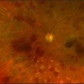
 Ocular ischemic syndrome
Jan 28 2018 by Alex H. Rubowitz, MD
A 65-year-old man with diabetes, presented with the typical round hemorrhages of OIS, as well as rubeosis iridis, and high intraocular pressure. Workup revealed carotid artery stenosis. Photos were taken just prior to his second laser PRP treatment.
Photographer: Lilach, Meir Hosopital Eye Clinic
Imaging device: Optos California
Condition/keywords: ocular ischemic syndrome
-
 OIS-Optos-photo-1
Jan 28 2018 by Alex H. Rubowitz, MD
A 65-year-old man with diabetes, presented with the typical round hemorrhages of OIS, as well as rubeosis iridis, and high intraocular pressure. Workup revealed carotid artery stenosis. Photos were taken just prior to his second laser PRP treatment.
Photographer: Lilach, Meir Hosopital Eye Clinic
Imaging device: Optos California
Condition/keywords: ocular ischemic syndrome
-
 Ocular Ischemic Syndrome
Jan 27 2018 by Alex H. Rubowitz, MD
A 65-year-old male with known diabetic retinopathy presented with iris rubeosis and neovascular glaucoma in his left eye. Photo was taken prior to his secind laser PRP session in the eye, and shows typical round hemorrhages of OIS. A systemic workup revealed severe carotid stenosis.
Photographer: Lilach, Meir Hospital Retina Clinic
Imaging device: Optos California
Condition/keywords: ocular ischemic syndrome
-
 Ocular Ischemic Symdrome
Jan 27 2018 by Alex H. Rubowitz, MD
A 65-year-old male with known diabetic retinopathy presented with iris rubeosis and neovascular glaucoma in his left eye. Photo was taken prior to his secind laser PRP session in the eye, and shows typical round hemorrhages of OIS. A systemic workup revealed severe carotid stenosis.
Photographer: Lilach, Meir Hospital Retina Clinic
Imaging device: Optos California
Condition/keywords: ocular ischemic syndrome
-
 Ocular Ischemic Syndrome With Neovascularization Due to Left Cartoid Artery Occlusion
Jan 8 2015 by Connie J Chen, MD
Fundus photo of a 66-year-old female with insulin dependent diabetes. She presented with eye pain due to neovascular glaucoma and angiography demonstrates extensive non-perfusion in the left eye with leakage from neovascularization of the disc. The right eye is completely perfused with no neovascular changes
Photographer: David Emmert
Imaging device: Optos Widefield Angiography
Condition/keywords: non-perfusion, ocular ischemic syndrome
-
 Ocular Ischemia From Left Carotid Artery Occlusion
Jan 8 2015 by Connie J Chen, MD
Fundus photo of a 66-year-old female with insulin dependent diabetes. She presented with eye pain due to neovascular glaucoma and angiography demonstrates extensive non-perfusion in the left eye with leakage from neovascularization of the disc. The right eye is completely perfused with no neovascular changes
Photographer: David Emmert
Imaging device: Optos Widefield Angiography
Condition/keywords: non-perfusion, ocular ischemic syndrome
-
 Ocular Ischemia
Dec 12 2014 by David Callanan, MD
66-year-old white male, ocular ischemia.
Condition/keywords: ocular ischemic syndrome
-
 Ocular Ischemia
Dec 12 2014 by David Callanan, MD
66-year-old white male, ocular ischemia.
Condition/keywords: ocular ischemic syndrome
-
 Ocular Ischemia
Dec 12 2014 by David Callanan, MD
66-year-old white male, ocular ischemia.
Condition/keywords: ocular ischemic syndrome
-
 Ocular Ischemia
Dec 12 2014 by David Callanan, MD
66-year-old white male, ocular ischemia.
Condition/keywords: ocular ischemic syndrome
-
 Ocular Ischemia
Dec 12 2014 by David Callanan, MD
66-year-old white male, ocular ischemia.
Condition/keywords: ocular ischemic syndrome
-
 Ocular Ischemia
Dec 12 2014 by David Callanan, MD
66-year-old white male, ocular ischemia.
Condition/keywords: ocular ischemic syndrome
-
 Ocular Ischemia
Dec 12 2014 by David Callanan, MD
66-year-old white male, ocular ischemia.
Condition/keywords: ocular ischemic syndrome
 Loading…