Search results (130 results)
-
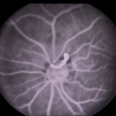
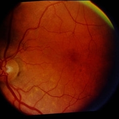
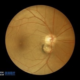 Double disc sign
Oct 13 2022 by Vaibhavi Noticewala, M S Ophthalmology, FVRS
Double disc sign Doubling of the optic disc is rare and can manifest as true or pseudo doubling. Duke-Elder describes duplication of the optic disc as a rare anomaly wherein two discs, each provided with retinal vessels are seen in an otherwise normal eye. Rare cases of true duplication of optic discs with separation of optic nerve into two or more strands have been reported, based either on incidental necropsy findings, demonstration of two optic foramina in the same orbit on x ray, or angioscotomas as indirect evidence of the existence of double optic nerves. Pseudo doubling of the optic discs caused by lesions such as optic disc coloboma, peripapillary chorioretinal coloboma, or inflammatory foci are more common. Our case had Ipsilateral isolated ectatic peripapillary chorioretinal coloboma simulating double optic discs.
Photographer: Priyal Mistry
Condition/keywords: Pseudoduplication of optic disc
-
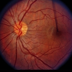
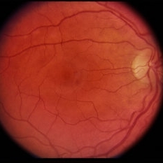
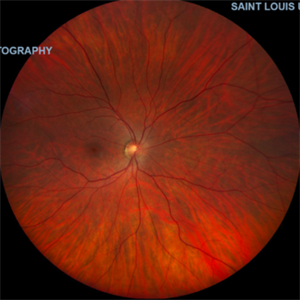 Normal Fundus Photo, OD in Pt. with Myelinated NFL, OS
Oct 27 2021 by Charles Hurth
Fundus photograph of a 35-year-old woman with myelinated nerve fiber layer in her left eye who presented to the adult strabismus clinic with exotropia of her left eye.
Photographer: Charles Hurth, IV, DO, Saint Louis University
Condition/keywords: normal eye
-
 Puzzle Retinitis
Jan 20 2021 by Jamin S. Brown, MD
Puzzle artifact after imaging on a smaller field of view with blue light autofluorescence.
Photographer: Stefanie Palmer CRA, Retina Vitreous Surgeons of CNY
Condition/keywords: autofluorescence imaging, normal eye
-
 Left -Anterior Segment
Aug 10 2020 by RITESH VERMA
Normal anterior segment of the left eye.
Photographer: Dr. Ritesh Verma, Regional institute of Ophthalmology, Rohtak, Haryana, India
Imaging device: CR-2AF CANON
Condition/keywords: anterior segment, normal eye
-
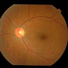
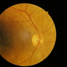
 Normal Fundus
Mar 26 2019 by Gary R. Cook, MD, FACS
Normal fundus photograph OS; VA= 20/20.
Imaging device: Topcon VT-50
Condition/keywords: normal eye
-
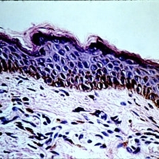 Slide 5-1
Feb 20 2019 by Lancaster Course in Ophthalmology
Normal lid skin from a darkly pigmented individual.
Condition/keywords: normal eye
-
 Normal Eye
-
 Preretinal Vascular Loop
Feb 20 2015 by H. Michael Lambert, MD
Vascular loop on the optic nerve, mid-phase fluorescein angiogram. 31-year-old black male, else normal eye exam. Father, brother and daughter also had vascular abnormalities.
Condition/keywords: dominantly inherited, optic nerve, vascular loop
-
 Preretinal Vascular Loop
Feb 20 2015 by H. Michael Lambert, MD
Vascular loop on the optic nerve, early fluorescein angiogram. 29-year-old black male, else normal eye exam. Father, sister and nephew also had vascular abnormalities.
Condition/keywords: dominantly inherited, optic nerve, vascular loop
-
 Preretinal Vascular Loop
Feb 20 2015 by H. Michael Lambert, MD
Vascular loop on the optic nerve, mid-phase fluorescein angiogram. 29-year-old black male, else normal eye exam. Father, sister and nephew also had vascular abnormalities.
Condition/keywords: dominantly inherited, optic nerve, vascular loop
-
 Preretinal Vascular Loop
Feb 20 2015 by H. Michael Lambert, MD
Vascular loop on the optic nerve. 29-year-old black male, else normal eye exam. Father, sister and niece also had vascular abnormalities.
Condition/keywords: dominantly inherited, optic nerve, vascular loop
-
 Preretinal Vascular Loop
Feb 20 2015 by H. Michael Lambert, MD
Vascular loop on the optic nerve. 29-year-old black male, else normal eye exam. Father, sister and niece also had vascular abnormalities.
Condition/keywords: dominantly inherited, optic nerve, vascular loop
-
 Preretinal Vascular Loop
Feb 20 2015 by H. Michael Lambert, MD
29-year-old black male, else normal eye exam. Father, sister and niece also had vascular abnormalities.
Condition/keywords: dominantly inherited, optic nerve, vascular loop
-
 Preretinal Vascular Loop
Feb 20 2015 by H. Michael Lambert, MD
Vascular loop on the optic nerve. 29-year-old black male, else normal eye exam. Father, sister and niece also had vascular abnormalities.
Condition/keywords: dominantly inherited, optic nerve, vascular loop
-
 Preretinal Vascular Loop
Feb 20 2015 by H. Michael Lambert, MD
Vascular loop on the optic nerve. 31-year-old black female, else normal eye exam. Father, brother and daughter also had vascular abnormalities.
Condition/keywords: dominantly inherited, optic nerve, vascular loop
-
 Preretinal Vascular Loop
Feb 20 2015 by H. Michael Lambert, MD
Vascular loop on the optic nerve. 31-year-old black female, else normal eye exam. Father, brother and daughter also had vascular abnormalities.
Condition/keywords: dominantly inherited, optic nerve, vascular loop
-
 Preretinal Vascular Loop
Feb 20 2015 by H. Michael Lambert, MD
Vascular loop on the optic nerve. 31-year-old black female, else normal eye exam. Father, brother and daughter also had vascular abnormalities.
Condition/keywords: dominantly inherited, optic nerve
-
 Normal Left Eye
-
 Normal Fundus
Feb 12 2015 by H. Michael Lambert, MD
Normal fundus, left eye.
Condition/keywords: left eye, normal eye
-
 Normal Appearing Fundus
Jan 30 2015 by H. Michael Lambert, MD
Normal appearing fundus, right eye.
Condition/keywords: normal eye
-
 Toxocara
Jan 7 2015 by H. Michael Lambert, MD
Left eye normal macula.
Condition/keywords: normal eye, toxocara canis
-
 Normal
Jan 7 2015 by H. Michael Lambert, MD
Color photograph of normal fundus.
Condition/keywords: normal eye, normal retina
-
 Normal Fundus
-
 Normal Fundus
-
 Normal F/A
 Loading…