Search results (54 results)
-
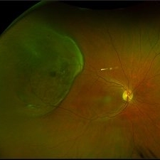
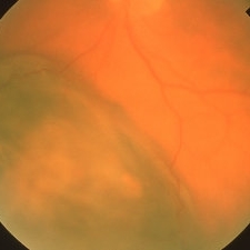
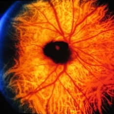
 RPE Rip s/p Brachytherapy for Malignant Melanoma
Jun 20 2025 by Virginia Gebhart
77 year old female with regressing tumor 4 months s/p brachytherapy. RPE rip at inferior edge of lesion.
Photographer: Virginia Gebhart, Retina Consultants of Carolina
Imaging device: Optos California
Condition/keywords: brachytherapy, choroidal melanoma, RPE Rip
-
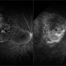 FA Malignant Melanoma
Aug 18 2023 by Angela Rico
A 48 year old female who presented with peripheral flashes and floaters , 1 month duration Fluorescein angiogram of Malignant Melanoma. Picture on left- Timer 00:08:163 Picture on right- Timer 04:08:92
Photographer: Angela Rico M.D.
Condition/keywords: malignant melanoma
-
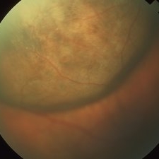
 Malignant Melanoma
Aug 18 2023 by Angela Rico
Fundus photograph of a 48 year old female with history of peripheral flashes and floaters OD for one month
Photographer: Angela Rico M.D.
Condition/keywords: intraocular tumor, malignant melanoma
-
 Presumed Malignant Melanoma
Jul 16 2021 by Rohit Ross Lakhanpal, MD FACS FASRS
Optos image of 59-year-old woman with complaint of dark shadow in her vision noted by closing her right eye.
Imaging device: Optos Camera
Condition/keywords: malignant neoplasm of eye
-
 Radiation Retinopathy Post Plaque I-125 Brachytherapy Treating Choroidal Melanoma
Apr 23 2021 by Sophia El Hamichi, MD
A 70-year-old-female with a history of choroidal melanoma treated with I-125 brachytherapy OS. She presented with radiation retinopathy in that eye.
Imaging device: Optos
Condition/keywords: I-125 brachytherapy, malignant melanoma, radiation retinopathy, ultra-wide field imaging
-
 Enucleated Eye with Malignant Melamoma
Apr 13 2020 by Sophia El Hamichi, MD
A 84-year-old female with advanced malignant melanoma of the her left eye requiring enucleation.
Photographer: Belinda Rodriguez, Murray Ocular Oncology and Retina, Miami
Condition/keywords: enucleation, gross pathology, malignant melanoma
-
 Choroidal Malignant Melanoma
Apr 26 2019 by Carissa Hurdstrom
Malignant choroidal melanoma
Photographer: Carissa Hurdstrom
Imaging device: Topcon 50DX
Condition/keywords: color fundus photograph
-
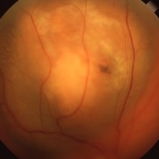
 Malignant Melanoma
Apr 1 2019 by Gary R. Cook, MD, FACS
Medium-size malignant melanoma with lipofuscin pigment.
Imaging device: Topcon VT-50
Condition/keywords: malignant melanoma
-
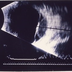 Malignant Melanoma
Apr 1 2019 by Gary R. Cook, MD, FACS
B-scan image of the malignant melanoma in the left eye of an 81-year-old white male; the simultaneous A-scan shows a high initial spike with low to medium internal reflectivity; B-scan shows some tenting of the retina over the melanoma; V.A.= 20-25-1.
Condition/keywords: malignant melanoma
-
 Malignant Melanoma
Apr 1 2019 by Gary R. Cook, MD, FACS
81-year-old white male with malignant melanoma inferiorly OS; V.A.= 20/25-1.
Imaging device: Topcon VT-50
Condition/keywords: malignant melanoma
-
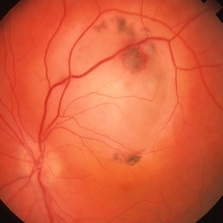
 Malignant Melanoma
Apr 1 2019 by Gary R. Cook, MD, FACS
87-year-old white male with a small malignant melanoma measuring 7 x 5 x 2.3mm by photography and ultrasonography; lipofuscin pigment is visible on its surface; V.A.= 20/200
Imaging device: Topcon VT-50
Condition/keywords: lipofuscin, malignant melanoma
-
 Ciliochoroidal Melanoma
Apr 1 2019 by Gary R. Cook, MD, FACS
34-year-old white male with a large ciliochoroidal melanoma superiorly; V.A.= 20/50-2; the eye was enucleated.
Imaging device: Topcon VT-50
Condition/keywords: ciliary body mass, malignant melanoma, malignant neoplasm of eye, melanoma
-
 Malignant Melanoma
Apr 1 2019 by Gary R. Cook, MD, FACS
56-year-old white male with a medium-size malignant melanoma OD; V.A. = 20/200.
Imaging device: Topcon VT-50
Condition/keywords: malignant melanoma
-
 Amelanotic Malignant Melanoma
Apr 1 2019 by Gary R. Cook, MD, FACS
White female with medium-sized, largely amelanotic malignant melanoma in posterior pole OS.
Imaging device: Topcon VT-50
Condition/keywords: malignant melanoma
-
 Slide 14-18
Mar 4 2019 by Lancaster Course in Ophthalmology
The most convincing evidence for the relationship of uveal nevi to malignant melanoma is the histologic observation of presumed nevus cells in association with ocular malignant melanomas in enucleated eyes.
Condition/keywords: malignant melanoma, melanoma
-
 Slide 11-40
Feb 26 2019 by Lancaster Course in Ophthalmology
Clinical appearance of a melanocytoma of the optic disk. This tumor is more common in blacks and in persons with congenital melanosis. It should not be confused with malignant melanoma.
Condition/keywords: melanocytoma, optic disc
-
 Slide 2-30
Feb 19 2019 by Lancaster Course in Ophthalmology
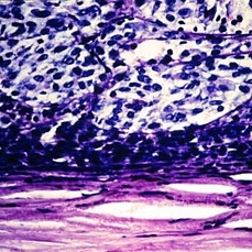
Malignant melanoma of the choroid. Smaller, deep blue cells over the tumor are lymphocytes.
Condition/keywords: choroid, lymphocytes, melanoma
-
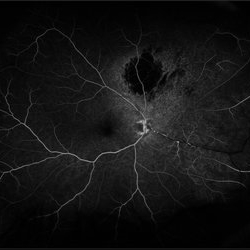 Radiation Retinopathy resulting in Retinal Vascular Occlusive Disease
Sep 11 2018 by Olivia Rainey
Ultra-wide field fluorescein angiography of a 57-year-old male s/p I-125 brachytherapy for malignant melanoma affecting his right eye. The patient’s radiation retinopathy has resulted in retinal vascular occlusive disease and optic nerve edema.
Photographer: Olivia Rainey
Imaging device: Optos
Condition/keywords: branch retinal vein occlusion (BRVO), fluorescein angiogram (FA), I-125 brachytherapy, malignant melanoma, optic disc edema, Optos, radiation retinopathy, ultra-wide field imaging
-
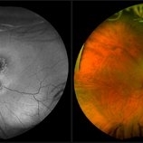 Malignant Melanoma
Sep 11 2018 by Olivia Rainey
Ultra-wide field autofluorescence and pseudocolor montage of a 57-year-old male s/p I-125 brachytherapy for malignant melanoma affecting his right eye. The patient’s radiation retinopathy has resulted in retinal vascular occlusive disease and optic nerve edema.
Photographer: Olivia Rainey
Imaging device: Optos
Condition/keywords: branch retinal vein occlusion (BRVO), fundus autofluorescence (FAF), I-125 brachytherapy, malignant melanoma, montage, Optos, pseudocolor, radiation retinopathy, ultra-wide field imaging
-
 Malignant Melanoma
Apr 11 2016 by Kathy Karsten, COT
Fundus photo montage of a malignant melanoma in a 70-year-old woman.
Photographer: KATHY KARSTEN, COT
Imaging device: TOPCON TRC 50 DX
Condition/keywords: malignant melanoma
-
 Collar Button Choroidal Melanoma
Oct 25 2015 by Dwain G. Fuller, MD, JD
Fundus photograph of collar button choroidal melanoma with associated serous retinal detachment and pre-retinal hemorrhages.
Condition/keywords: collar button, malignant melanoma
-
 Collar Button Choroidal Melanoma
Oct 25 2015 by Dwain G. Fuller, MD, JD
Fundus photograph of collar button choroidal melanoma.
Imaging device: Optos
Condition/keywords: collar button, malignant melanoma
-
 Collar Button Choroidal Melanoma
Oct 25 2015 by Dwain G. Fuller, MD, JD
Ultrasonographic image of collar button choroidal melanoma with associated serous retinal detachment.
Photographer: Jana Sierocki
Condition/keywords: collar button, malignant melanoma
-
 Collar Button Choroidal Melanoma
Oct 25 2015 by Dwain G. Fuller, MD, JD
Fundus photograph of large collar button choroidal melanoma.
Condition/keywords: collar button, malignant melanoma
-
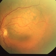
 Malignant Melanoma
Jul 8 2015 by Kathy Karsten, COT
Fundus photograph of 70-year-old woman presenting with obscured vision in the right eye. Diagnosed with malignant melanoma. Patient underwent subsequent brachytherapy.
Photographer: Kathy Karsten, COT
Imaging device: Topcon TRC-50DX
 Loading…