Search results (8 results)
-
 Macula On Retinal Detachment
Jul 5 2024 by Zach Seim
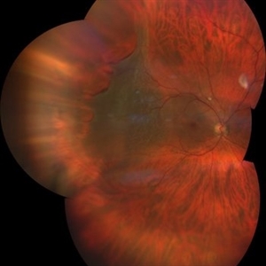
This is an Optos fundus photo of a 67 year old female with a Macula On Retinal Detachment. Patient presented with VA DCC 20/40-1.
Photographer: Zach Seim
Imaging device: Optos California
Condition/keywords: macula on, Optos, OPTOS CALIFORNIA, right eye
-
 Retinal Detachment
Jan 31 2024 by Cuitláhuac del Moral Herrera
69 year old male presented with a week long scotoma on his right eye. A macula ON retinal detachment with a “spokes-wheel” giant tear was noted upon examination. We performed a phaco-vit with good results.
Photographer: Manuela Franco Sánchez, Asociación para Evitar la Ceguera en México. Cuitláhuac del Moral Herrera, Asociación para Evitar la Ceguera en México.
Imaging device: ZEISS CLARUS 700
Condition/keywords: macula on, Spokes wheel
-
 Macula on Retinal Detachment with large Horseshoe Tear
Apr 26 2023 by Kelli Nyenhuis
Optos photograph of a 61-year-old male with a macula on retinal detachment and large horseshoe tear. Patient had no visual changes.
Photographer: Kelli Nyenhuis, COA
Imaging device: Optos California
-
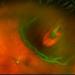
 Subretinal Migration of SF6 gas: Postop day 1
Jan 29 2022 by Raja Rami P Reddy, MD FRCS FASRS
67-year-old with a new Rhegmatogenous Retinal Detachment, macula on, with posterior retinal tear, superotemporally. Patient underwent 25 G vitrectomy, endo laser and SF6 gas injection and was advised prone position postoperatively. The image shows the appearance of the fundus on postoperative day 1 through the gas bubble . Patient was advised sitting position subsequently. Resolved completely over the next five days.
Photographer: Raja Rami Reddy P, neoretina eyecare institute
Imaging device: Optos, Daytona
Condition/keywords: Subretinal migration, sulphur hexafluoride gas
-
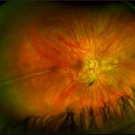 Prominent Long Ciliary Nerve
Jan 25 2022 by Kachelle Brown
Ultra-wide field photograph of a 48-year-old female with a prominent long ciliary nerve. Patient presented asymptomatic, and was referred for a macula on retinal detachment. Patient was diagnosed with high myopia and a posterior vitreous detachment, and the physician discussed increased risk of floaters, myopic degeneration and retinal detachment associated with high myopia. -24.00 prior to cataract surgery OU per patient.
Photographer: Kachelle Brown
Imaging device: Optos California
Condition/keywords: fundus photograph, high myopia, long ciliary nerve, optos, right eye, ultra-widefield image
-
 Macula ON Retinal Detachment
Feb 18 2021 by Omar Lezrek
Fundus mosaic of an 26-year-old woman with macula ON retinal detachment with 9 o'clock retina tear.
Photographer: Omar Lezrek MD
Imaging device: Eidon AF
Condition/keywords: retinal detachment with retinal defect
-
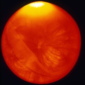 Giant Retinal Tear With Retinal Detachment, Macula On
Oct 2 2013 by Jerald A. Bovino, MD
This patient has a giant retinal tear with a retinal detachment. The macula is still attached.
Condition/keywords: macula, retinal tear
-
 Cystoid Macular Edema
Dec 12 2012 by Mallika Goyal, MD
Left eye of a patient with proliferative diabetic retinopathy and cystoid macular edema shows characteristic petalloid appearance at macula on fluorescein angiography.
Photographer: Mallika Goyal, MD, Apollo Hospitals, Hyderabad, India
Condition/keywords: cystoid macular edema (CME)
 Loading…