Search results (9 results)
-
 Dislocated Iol With Hypotony Maculopathy and Hemorrhagic Choroidal
Feb 9 2024 by Sandra R Montezuma, MD
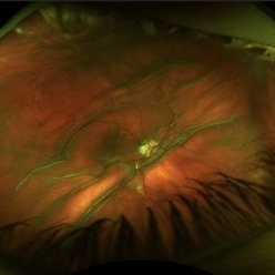
28 year old year-old male with history of congenital cataract of the right eye, s/p cataract extraction in 1999, s/p lens implant in 2011, presented with a dislocated IOL, hypotony, retina folds, hypotony maculopathy and hemorrhagic nasal choroidal after unsuccessful surgery to attempt remove the dislocated lens.
Photographer: Scott Baker, University of Minnesota
Condition/keywords: choroidals, dislocated posterior chamber intraocular lens (PCIOL), hypotony maculopathy, retina folds
-
 Hypotony Maculopathy
Nov 3 2023 by Matthew Dombrow, MD
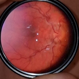
31 year old female 4 days s/p Ahmed Valve
Photographer: Cori Sturtevant, Connecticut Retina Consultants, Hamden, Connecticut
Imaging device: Optos - California
Condition/keywords: hypotony maculopathy
-
 Hypotony maculopathy
Mar 13 2023 by Pawel Kolman
20 y.o male with hypotony (4 mmHg) caused by cilliary body shutdown in setting of anterior uveitis.
Photographer: Pawel Kolman
Imaging device: Volk 20D and Samsung Galaxy S21
Condition/keywords: hypotony, hypotony maculopathy, uveitis
-
 Hypotony maculopathy
Feb 23 2023 by Kamal Kishore, MD, MBBS
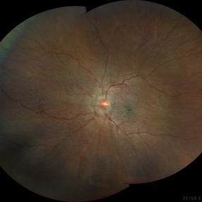
Ultrawide field fundus photograph of a 62-year-old male with hypotony following blunt ocular trauma
Photographer: Kim Grabill, COA, Illinois Retinal and Eye Associates, Peoria, IL, USA
Imaging device: Zeiss Clarus
Condition/keywords: hypotony maculopathy
-
 Hypotony Maculopathy
Jun 12 2022 by Pramod Kumar Suman, MBBS, MD
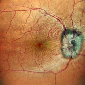
Fundus photograph of an 26-year-old male with retinal folds around the center of the fovea arranged in stellate pattern with optic disc edema.
Photographer: Pramod Kumar Suman, Retina Foundation, Ahmedabad
Condition/keywords: hypotony maculopathy
-
 Hypotonic Maculopathy Following Glaucoma Surgery
Mar 27 2021 by Deepak Bhojwani, MS
Fundus image of a 48-year-old female with recent history of glaucoma surgery showing edematous disc, diffuse hypotonic choroidal folds, prominent macular hypotonic folds and massive choroidal in mid-periphery.
Photographer: DEEPAK BHOJWANI; VISHAL PATEL FOR OCCURA EYE CARE
Condition/keywords: hypotony maculopathy
-
 Hypotony Maculopathy
Apr 1 2019 by Anfisa Ayalon, MD
Fundus autofluorescence image of 81-year-old male with right eye ocular hypotony due to leaking bleb. Note severe hypotony maculopathy, peripheral choroidal detachments, multiple chorioretinal folds.
Photographer: Anfisa Ayalon, MD., Meir Medical Center, Kfar Saba, Israel.
Imaging device: California, Optos 200 DTX
Condition/keywords: choroidal detachment, choroidal folds, fundus autofluorescence (FAF), hypotonous retinopathy, hypotony maculopathy
-
 Ocular Hypotony Due to Leaking Bleb
Apr 1 2019 by Anfisa Ayalon, MD
81-year-old male who had trabeculectomy in his right eye 4 years ago, presented to the emergency room with complains of decreased vision in that eye for two months. Slit-lamp examination showed cystic bleb with leakage, intraocular pressure was 0 MMHg. Fundus examination showed hypotony maculopathy, peripheral choroidal detachments, multiple chorioretinal folds with subretinal fluid.
Photographer: Anfisa Ayalon, MD., Meir Medical Center, Kfar Saba, Israel.
Imaging device: California, Optos 200 DTX
Condition/keywords: choroidal detachment, hypotonous retinopathy, hypotony maculopathy
-
 Hypotony Maculopathy
May 3 2018 by Alexandr Stepanov
Hypotony maculopathy.
Photographer: Alexandr Stepanov MD, PhD, FEBO, Faculty Hospital Hradec Kralove, Czech Republic
Condition/keywords: hypotony maculopathy
 Loading…