Search results (159 results)
-
 Hypertensive Retinopathy
Sep 16 2025 by Píndaro Alonso Cruz-Benitez
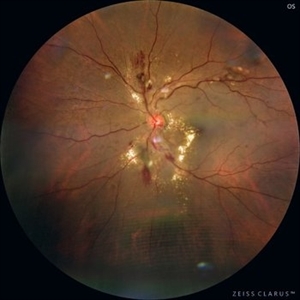
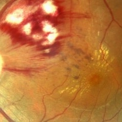
Hypertensive Retinopathy Stage 3 Male 55 year-old
Photographer: Pindaro Alonso Cruz Benitez
Condition/keywords: exudate, Haemorrhage, hypertensive retinopathy
-
 Hyperthensive Retinopathy
Sep 16 2025 by Píndaro Alonso Cruz-Benitez
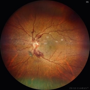
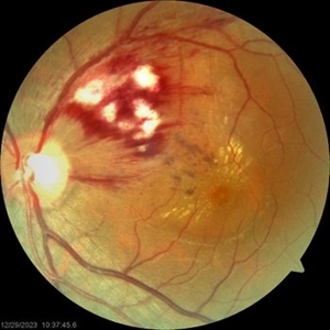
55 year-old male Hyperthensive Retinopathy Stage IV Papilledema
Photographer: Píndaro Alonso Cruz-Benítez
Condition/keywords: hypertensive retinopathy, papilledema, Retina
-
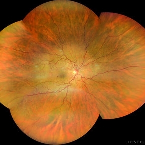
 Hypertensive Retinopathy
Jun 4 2025 by Paulina Araujo
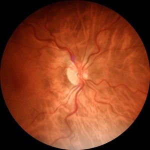
The 55-degree central fundus photograph of the right eye reveals vascular tortuosity, generalized arteriolar narrowing with a vein-to-artery ratio of 3:1, along with Guist and Bonnet signs.
Photographer: Paulina D.Araujo Martínez, Asociación para Evitar la Ceguera en México I.A.P., Hospital Dr Luis Sánchez Bulnes.
Condition/keywords: hypertensive retinopathy
-
 Hypertensive Retinopathy
May 26 2025 by César Adrián Gómez Valdivia, MD
Fundus photograph of a 62 year-old woman with history of untreated hypertension and chronic kidney disease. Findings were bilateral.
Photographer: @eyemissu2
Imaging device: OPTOS
Condition/keywords: hypertensive retinopathy
-
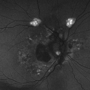 Astrocytic Hamartoma
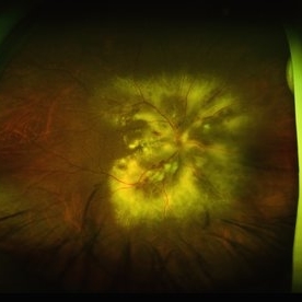
Feb 27 2025 by Daniel Davis, OCT-C
Fundus autofluorescence photo of 55-year-old female with astrocytic hamartoma in association with tuberous sclerosis. No treatment options available, benign. Other findings include; Posterior Vitreous Detachment, Vitreous Hemorrhage, Hereditary Retinal Dystrophy, Vitreous Opacities, Hypertensive Retinopathy.
Photographer: Daniel Davis, OCT-C
Imaging device: Optos California
Condition/keywords: astrocytic hamartoma, fundus autofluorescence (FAF)
-
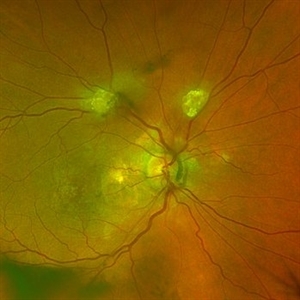 Astrocytic Hamartoma
Feb 27 2025 by Daniel Davis, OCT-C
Color fundus photo of 55-year-old female with Astrocytic Hamartoma in association with tuberous sclerosis. No treatment options available, benign. Other findings include; Posterior Vitreous Detachment, Vitreous Hemorrhage, Hereditary Retinal Dystrophy, Vitreous Opacities, Hypertensive Retinopathy.
Photographer: Daniel Davis, OCT-C
Imaging device: Optos California
Condition/keywords: color fundus photograph
-
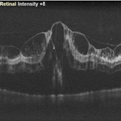 Garland of Superficial Infarct on OCT
Oct 29 2024 by Prashant K Bawankule, M.S.
OCT of 78 year-old hypertensive male with multiple soft exudates over posterior pole on clinical examination.
Photographer: Prashant Bawankule, Sarakshi Netralaya, Nagpur, Maharashtra , India
Imaging device: Mirante ( by Nidek)
Condition/keywords: Hypertensive retinopathy, Superficial infarcts
-
 Hypertensive Retinopathy
Oct 27 2024 by César Adrián Gómez Valdivia, MD
Fundus photograph of a 62 year-old woman with history of untreated hypertension and chronic kidney disease. Findings were bilateral.
Photographer: @eyemissu2
Imaging device: TOPCON TRC-50DX
Condition/keywords: hypertensive retinopathy
-
 Hypertensive Retinopathy
Sep 8 2024 by Cesar Augusto Rocha Rojas, MD
Fundus photograph of a 27-year-old male with hypertensive emergency secondary to chronic kidney disease.
Photographer: Cesar Augusto Rocha Rojas, Hospital General de Zona #20, Instituto Mexicano del Seguro Social
Imaging device: Smartphone, Pan Retinal 2.2 Lens
Condition/keywords: hypertensive retinopathy
-
 Hypertensive Retinopathy
May 1 2024 by Marco Antonio Sauza
36 year old male with uncontrolled systemic hypertension.
Photographer: MARCO SAUZA CASTILLEJOS
Imaging device: VISUCAM ZEISS
Condition/keywords: choroidal infarction, hypertensive retinopathy, macular star
-
 Hypertensive Retinopathy Grade IV
May 1 2024 by Marco Antonio Sauza
Fundus photograph of an 36-year-old male with uncontrolled systemic hypertension, >200/100mmhg, presenting decreased vision in the left eye.
Photographer: MARCO SAUZA CASTILLEJOS
Imaging device: VISUCAM ZEISS
Condition/keywords: choroidal infarction, hypertensive retinopathy, macular star
-
 Hypertensive Retinopathy
Apr 21 2024 by César Adrián Gómez Valdivia, MD
Hypertensive Retinopathy
Photographer: Erika Paulina Ornelas Cazares
Imaging device: TOPCON TRC-50DX
Condition/keywords: hypertensive retinopathy
-
 Central Retinal Artery Occlusion
Mar 11 2024 by Dr.Pavithra Subramanian
A 51 year old male with defective vision in right eye for past 4days.On examination RE RAPD present and Fundus examination found to be Right eye Central retinal artery occlusion with Grade 4 Hypertensive Retinopathy.
Photographer: Dr Pavithra Subramanian
Condition/keywords: central retinal artery occlusion (CRAO), malignant hypertension
-
 Malignant Hypertension
Sep 28 2023 by ANKIT JAIN
Fundus photograph of RE of a 13 year old male with malignant hypertension
Photographer: Dr. Ankit Jain
Imaging device: Mirante
Condition/keywords: hypertensive retinopathy, malignant hypertension
-
 Malignant Hypertension
Sep 28 2023 by ANKIT JAIN
Fundus photograph of LE of a 13 year old male with malignant hypertension
Photographer: Dr. Ankit Jain
Imaging device: Mirante
Condition/keywords: hypertensive retinopathy, malignant hypertension
-
 Malignant Hypertension
Sep 28 2023 by ANKIT JAIN
Widefield photograph of LE of a 13 year old male with malignant hypertension
Photographer: Dr. Ankit Jain
Imaging device: Mirante
Condition/keywords: hypertensive retinopathy, malignant hypertension
-
 RE WIDEFIELD FUNDUS PHOTOGRAPH OF HYPERTENSIVE RETINOPATHY GRADE IV
Sep 27 2023 by ANKIT JAIN
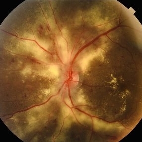
WIDEFIELD IMAGE OF RE OF 14 YEARS OLD MALE RECENTLY DIAGNOSED WITH MALIGNANT HYPERTENSION SHOWING MACULAR STAR, DISC EDEMA IN A CASE OF HYPERTENSIVE RETINOPATHY GRADE IV.
Photographer: DR ANKIT JAIN
Imaging device: MIRANTE
Condition/keywords: Disc Edema, hypertensive retinopathy, macular star
-
 LE WIDEFIELD FUNDUS PHOTOGRAPH OF HYPERTENSIVE RETINOPATHY GRADE IV
Sep 27 2023 by ANKIT JAIN
WIDEFIELD IMAGE OF RE OF 14 YEARS OLD MALE RECENTLY DIAGNOSED WITH MALIGNANT HYPERTENSION SHOWING MACULAR STAR, DISC EDEMA IN A CASE OF HYPERTENSIVE RETINOPATHY GRADE IV.
Photographer: DR ANKIT JAIN
Imaging device: MIRANTE
Condition/keywords: Disc Edema, hypertensive retinopathy, macular star
-
 Hypertensive Retinopathy
Sep 12 2023 by Ben Serar
Fundus photograph of RE showing Disc pallor and arteriolar attenuation, with dot-blot and flame-shaped haemorrhages at the posterior pole in a case of Hypertensive Retinopathy.
Condition/keywords: arteriolar attenuation, hypertensive retinopathy, pale disc
-
 Hypertensive Retinopathy
Sep 12 2023 by Ben Serar
Fundus photograph of LE showing Disc edema with optic disc pallor, hard exudates with dot-blot haemorrhages at the macula ,along with arteriolar attenuation, in a case of Hypertensive retinopathy.
Condition/keywords: arteriolar attenuation, disc edema, Hard exudates, hypertensive retinopathy
-
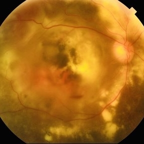
 Siegrist Streaks and Hypertensive Choroidopathy
Jul 29 2023 by Júlio Costa Almeida, MD
Fundus photograph of a 68-year-old woman with hypertensive retinopathy, choroidopathy and the classic Siegrist streaks.
Imaging device: Nonmyd 8s by Kowa
Condition/keywords: hypertensive choroidopathy, hypertensive retinopathy, Siegrist Streaks
-
 Diabetic Retinopahty
Nov 2 2022 by pedro fernandes souza neto
Fundus photograph of a 40-year-old man with diabetes and hypertension shows hard exudates, difuse intraretinal hemorrhages and splinter hemorrhages.
Photographer: Pedro Fernandes, Universidade Federal da Bahia, Brazil.
Condition/keywords: diabetic mellitus, hypertensive retinopathy, retinopathy
-
 Impending-CRVO
Feb 20 2022 by Vishal Gupta, MBBS, MS
Impending CRVO in a 54 year old hypertensive female patient with Collaterals and Grade 2 Chronic Hypertensive retinopathy.
Photographer: Dr Shobhit Chawla, Prakash Netra Kendr, Lucknow, UP, INDIA
Imaging device: Zeiss Clarus 500
Condition/keywords: central retinal vein occlusion (CRVO), collaterals, dilated tortuous vessels, hypertensive retinopathy
-
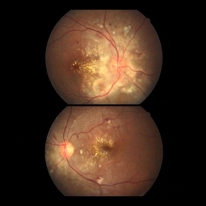 Hypertensive Retinopathy
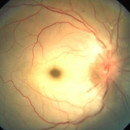
Jan 8 2022 by Gayathri Mohan
Color fundus image of both eyes showing a resolving hypertensive retinopathy, taken one month post episode of malignant hypertension.
Photographer: Dr Gayathri Mohan
Imaging device: Canon
Condition/keywords: hypertensive retinopathy, malignant hypertension
-
 Malignant-Hypertensive-Retinopathy-2
Nov 9 2021 by VIRAL SHAH
Disc edema and macular star in malignant hypertensive patient diagnosed with pheochromocytoma.
Photographer: VIRAL SHAH, NETRALOK RETINA CLINIC, AHMEDABAD
Condition/keywords: disc edema, hypertensive retinopathy, macular star
 Loading…