Search results (131 results)
-
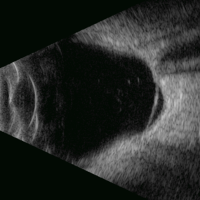
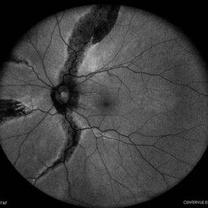
 RRD in Posterior Staphyloma
May 21 2025 by Gustavo Uriel Fonseca Aguirre
This B-mode axial ultrasound scan of a highly myopic eye demonstrates a prominent posterior staphyloma with an associated inferior retinal detachment sparing the macular region.
Photographer: Gustavo U. Fonseca Aguirre, Hospital Conde de Valenciana, Ciudad de México
Condition/keywords: high myopia, posterior staphyloma, Retina detachment
-
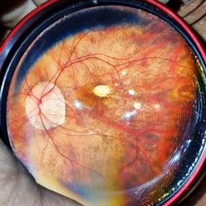
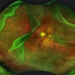 Giant Retinal Tear with Multiple Retinal Breaks
Apr 21 2025 by Hrishikesh Naik, MS
A 28 year old high myope with retinal detachment associated with a supero-temporal giant retinal tear in addition to multiple peripheral horseshoe tears and an additional supero-nasal retinal tear.
Photographer: Hrishikesh Naik
Imaging device: Optos Daytona
Condition/keywords: giant retinal tear, High Myopia, horseshoe tear, retinal break, retinal detachment
-
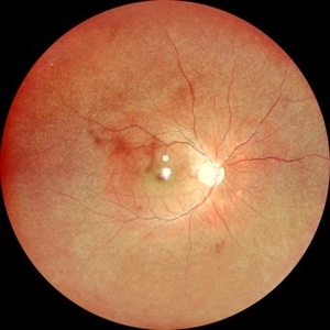
 Myelinated Nerve Fibers
Apr 18 2025 by DR Rohit Gupta
The **myelinated nerve fibers of the optic disc** (also known as **medullated nerve fibers**) are retinal nerve fibers that retain their myelin sheath as they pass through the optic nerve head. Normally, retinal nerve fibers are unmyelinated to allow for light transparency, but in some cases, myelination extends anteriorly into the retina, appearing as a striking white, feathery patch on the optic disc or peripapillary retina. ### **Key Features:** 1. **Appearance:** - Dense, white, striated patches with feathery edges. - Typically located at the superior or inferior pole of the optic disc. - May obscure retinal vessels underneath. 2. **Clinical Significance:** - Usually **benign** and asymptomatic. - **Congenital** (present at birth or early childhood). - Rarely associated with **visual field defects** (e.g., scotomas corresponding to the area of myelination). - Occasionally linked with **high myopia** or **amblyopia** if extensive. 3. **Pathophysiology:** - Failure of oligodendrocytes or Schwann cells to stop myelination at the lamina cribrosa. - Normally, myelination stops at the optic nerve head, but in this condition, it extends into the retina. 4. **Diagnosis:** - **Fundoscopy:** Classic white, feathery appearance. - **Optical Coherence Tomography (OCT):** Shows thickened retinal nerve fiber layer (RNFL). - **Visual Field Testing:** May detect defects if large. 5. **Differential Diagnosis:** - Optic disc edema - Cotton wool spots - Retinoblastoma (rarely, but must be ruled out in children) 6. **Management:** - No treatment required if asymptomatic. - Monitor for amblyopia in children. - Rare cases with significant visual impairment may need further evaluation. ### **Fun Fact:** Myelinated nerve fibers are seen in **~0.5-1%** of the population and are usually an incidental finding.
Photographer: Dr Rohit gupta
Imaging device: Samsung S21
Condition/keywords: Medulated Nerve fibre, Medullated Nerve fibres, myelinated nerve fibers, Myelinated Nerve Fibres, optic disc drusen
-
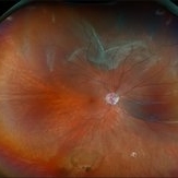
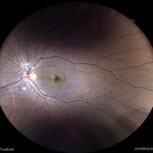
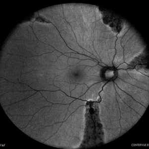
 Retinal Detachment Associated With a Posterior Staphyloma
Apr 9 2025 by Gustavo Uriel Fonseca Aguirre
B-mode axial ultrasound scan of a highly myopic eye shows a posterior staphyloma with an associated macular hole-induced retinal detachment.
Photographer: Gustavo U. Fonseca Aguirre, Hospital Conde de Valenciana, Ciudad de México
Condition/keywords: high myopia, posterior staphyloma, rhegmatogenous retinal detachment
-
 Retinitis Pigmentosa
Apr 9 2025 by Virginia Gebhart
35 year old female with stable sectoral RP and high myopia OU. RP has not progressed in either eye since initial visit in 2021. Will continue to observe. VA 20/20 OU
Photographer: Virginia Gebhart, Retina Consultants of Carolina
Imaging device: Optos California
Condition/keywords: high myopia, retinitis pigmentosa
-
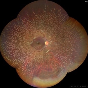
 Dome Shaped Macula
Jan 7 2025 by Jordyn Beckman
Autofluorescence photograph of 36 year old woman with Dome Shaped Macula with hypoautofluorescence and hyperautofluorescence centrally.
Photographer: Jordyn Beckman
Imaging device: California Optos
Condition/keywords: autoflorescence, convex protrusion, dome shaped macula, high myopia, hyperautofluorescent centrally, hypoautofluorescence
-
 Retinal Detachment with Multiple Breaks
Nov 4 2024 by Kimberly Wakester
Ultra-widefield Fundus photograph of an 18-year-old woman with a Retinal detachment with multiple breaks in the right eye. Patient has high Myopia in both eyes. Patient was treated with scleral buckle placement with cryo laser in the right eye and is doing we post operatively.
Photographer: Kimberly Wakester, COA
Imaging device: Optos
-
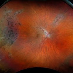
 Pathological Myopia
Sep 25 2024 by DR Rohit Gupta
Fundus photograph of a 28 year-old male having high myopia on fundus examination Degenerative changes are seen in retina suggestive of pathological myopia.
Photographer: Dr Rohit gupta
Imaging device: Samsung S21
Condition/keywords: choroidal degeneration, degeneration of optic disc, lacquer cracks, myopia, Myopia macular degeneration CNVM foster fuch spot, pathologic myopia, staphyloma
-
 Subclinical RD in Retinitis Pigmentosa
May 22 2024 by Tejaswita Verma
Fundus photograph of the right eye of a 6 year old male with subclinical retinal detachment with macula off in case of retinitis pigmentosa with high myopia.
Photographer: DR. TEJASWITA VERMA
Imaging device: MIRANTE
Condition/keywords: high myopia, retinitis pigmentosa, subclinical detachment
-
 Subclinical RD in Retinitis Pigmentosa
May 22 2024 by Tejaswita Verma
Fundus photograph of the left eye of a 6 year old male with subclinical retinal detachment with macula off in a case of retinitis pigmentosa with high myopia.
Photographer: DR. TEJASWITA VERMA
Imaging device: MIRANTE
Condition/keywords: high myopia, retinitis pigmentosa, subclinical detachment
-
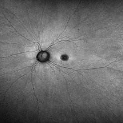
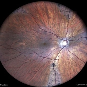
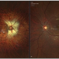 Right Myelinated Nerve Fibre, LE Normal
May 6 2024 by Anupama Janardhanan
Fundus photograph of a 24 year old male patient with Anisometropia and Right Eye High Myopia associated with Diffuse circumpapillary myelinated Nerve Fibre and Left Eye normal fundus.
Photographer: Dr. Anupama Janardhanan, Aravind Eye hospital, Tirunelveli, India
Imaging device: Heidelberg Spectralis
Condition/keywords: anisometropia, myelinated nerve fibers, myopic degeneration
-
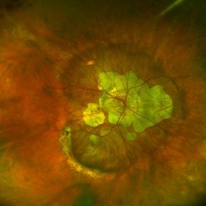
 Myopic Subretinal Neovascular Membrane
Apr 9 2024 by Akansha Sharma
Color fundus photograph of a 23 year old female with subretinal bleed in a case of high myopia.
Photographer: Dr. Akansha Sharma, Bharati Eye Hospital
Condition/keywords: myopic choroidal neovascularization (CNV), SRNVM, subretinal hemorrhage
-
 Lattice With Hole
Feb 6 2024 by Thirumalesh Mochi Basavaraj, MD
25 year old myopic patient with extensive lattice degeneration with multiple atrophic holes.
Photographer: Puttaswamy
Condition/keywords: atrophic hole, High myopia, lattice degeneration
-
 Lattice With Holes
Feb 6 2024 by Thirumalesh Mochi Basavaraj, MD
25 year old myopic patient with extensive lattice degeneration with multiple atrophic holes.
Photographer: Puttaswamy
Condition/keywords: atrophic retinal hole, High Myopia, peripheral lattice degeneration
-
 Benign Familial Fleck Retina
Dec 21 2023 by Vishal Agrawal, MD, FRCS,FACS,FASRS
10-year male with high myopia on examination revealed diffuse flecks distributed all over fundus in both eyes sparing macula. Inferior lattice with WWOP areas were also noted in right eye.
Photographer: Dr Ayushi
Imaging device: Clarus 700
Condition/keywords: fleck retinopathy, myopia
-
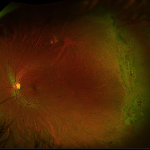
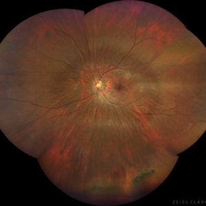
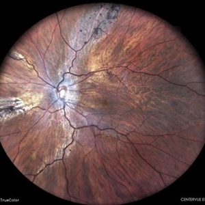
 Posterior staphyloma
Dec 20 2023 by Roger A. Goldberg, MD, MBA
Fundus photo of an 85-year-old woman with degenerative myopia and a large posterior staphyloma
Photographer: Mohan Zhou, Bay Area Retina Associates, Walnut Creek, CA
Imaging device: Optos
Condition/keywords: degenerative myopia, high myopia, posterior staphyloma
-
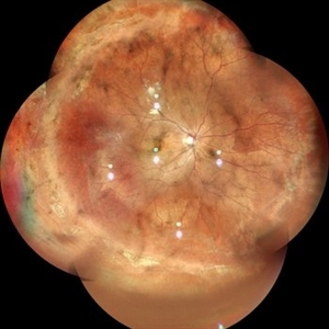
 High Myopia with Posterior staphyloma
Nov 7 2023 by Harsh Vardhan Singh, MS
27-year old with both eyes high myopia & posterior staphyloma with left eye peripheral lattice degeneration & white without pressure
Photographer: Harsh Vardhan Singh
Imaging device: Clarus 700
Condition/keywords: lattice degeneration, myopia, peripheral lattice degeneration, posterior staphylomaloma, white without pressure
-
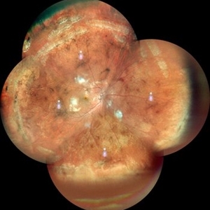
 Pathological Myopia
Oct 18 2023 by Anand Temkar
RE widefield CF montage of a 24 year old male with pathological myopia showing multiple lattice degenerations in periphery along with holes.
Photographer: Dr.Anand Temkar- Retina Foundation, Ahmedabad
Imaging device: Mirante
Condition/keywords: high myopia, holes, lattice degeneration
-
 Pathological Myopia
Oct 18 2023 by Anand Temkar
RE widefield color photo montage of a 25 years old male showing lattice degenerations in periphery in a case of pathological myopia.
Photographer: Dr.Anand Temkar- Retina Foundation, Ahmedabad
Imaging device: Mirante
Condition/keywords: high myopia, lattice degeneration
-
 LARGE OPERCULATED HOLE
Oct 16 2023 by ANKIT JAIN
Left Eye Montage of 50 years old male with high myopia with large operculated hole
Photographer: DR ANKIT JAIN
Imaging device: MIRANTE
Condition/keywords: High Myopia, myopia, operculated retinal hole
-
 Pigmented Paravenous Retinochoroidal Atrophy
Oct 1 2023 by Bruno DECAY, MD
Fundus photograph of a 43-year-old myopic man (right eye) Axial length: 27.67 mm
Photographer: Bruno DECAY, MD
Imaging device: iCARE EIDON confocal fundus imaging system (Ultra-Widefield Module)
Condition/keywords: Bilateral, High myopia, Pigmented Paravenous Retinochoroidal Atrophy
-
 Pigmented Paravenous Retinochoroidal Atrophy
Oct 1 2023 by Bruno DECAY, MD
Fundus photograph (autofluorescence) of a 43-year-old myopic man (right eye) Axial length: 27.67 mm
Photographer: Bruno DECAY, MD
Imaging device: iCARE EIDON confocal fundus imaging system (Ultra-Widefield Module)
Condition/keywords: Bilateral, High Myopia, Pigmented Paravenous Retinochoroidal Atrophy
-
 Pigmented Paravenous Retinochoroidal Atrophy
Oct 1 2023 by Bruno DECAY, MD
Fundus photograph of a 43-year-old myopic man (left eye) Axial length: 29.32 mm
Photographer: Bruno DECAY, MD
Imaging device: iCARE EIDON confocal fundus imaging system (Ultra-Widefield Module)
Condition/keywords: Bilateral, High myopia, Pigmented Paravenous Retinochoroidal Atrophy
-
 Pigmented Paravenous Retinochoroidal Atrophy
Oct 1 2023 by Bruno DECAY, MD
Fundus photograph (autofluorescence) of a 43-year-old myopic man (left eye) Axial length: 29.32 mm
Photographer: Bruno DECAY, MD
Imaging device: iCARE EIDON confocal fundus imaging system (Ultra-Widefield Module)
Condition/keywords: Bilateral, High myopia
-
 Myelinated Nerve Fibres in left eye with old tributary vein occlusion in left eye
Jul 18 2023 by Harsh Vardhan Singh, MS
55 year female with left eye amblyopia & high myopia with MNF and Right eye showed signs of old macular branch retinal vein occlusion
Photographer: Harsh Vardhan Singh, AIIMS, Guwahati
Imaging device: Zeiss Clarus 700
Condition/keywords: BRVO, macular branch retinal vein occlusion (BRVO), Medullated Nerve fibres, MNF, Myelinated Nerve Fibres, TRVO
 Loading…