Search results (11 results)
-
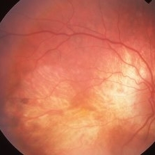
 Choroideremia
Sep 21 2022 by Zach Seim
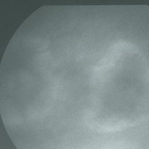
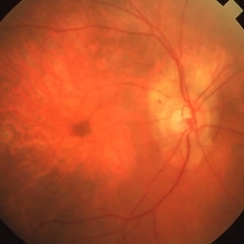
Ultra-widefield fundus photo of a 74 year old male presenting with severe vision loss beginning at age 55. Patient sought a second opinion with our office and was diagnosed with Choroideremia. Patient denies hearing loss, heart problems, balance issues, polydactyly, kidney problems, and dental problems. Patient reports that nobody in the family had blindness. Choroideremia is an X-linked chorioretinal dystrophy characterized by the diffuse, progressive degeneration of the retinal pigment epithelium (RPE), photoreceptors and choriocapillaris. It is caused by a mutation in the CHM gene.
Photographer: Zach Seim
Imaging device: Optos California
Condition/keywords: choroideremia, hereditary choroidal atrophy, hereditary retinal dystrophy, left eye, light perception, low vision, Optos, pseudocolor, ultra-wide field imaging
-
 Choroideremia
Sep 21 2022 by Zach Seim
Ultra-widefield fundus photo of a 74 year old male presenting with severe vision loss beginning at age 55. Patient sought a second opinion with our office and was diagnosed with Choroideremia. Patient denies hearing loss, heart problems, balance issues, polydactyly, kidney problems, and dental problems. Patient reports that nobody in the family had blindness. Choroideremia is an X-linked chorioretinal dystrophy characterized by the diffuse, progressive degeneration of the retinal pigment epithelium (RPE), photoreceptors and choriocapillaris. It is caused by a mutation in the CHM gene.
Photographer: Zach Seim
Imaging device: Optos California
Condition/keywords: choroideremia, hereditary choroidal atrophy, hereditary retinal dystrophy, Optos, pseudocolor, ultra-wide field imaging
-
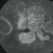
 Regional Choriocapillaris Atrophy
Jun 18 2019 by Gary R. Cook, MD, FACS
Late-phase (5 minutes) FA frame of the left eye of a 73-year-old white female with regional choriocapillaris atrophy showing light staining from intact choriocapillaris around the margins of the peripapillary and macular areas of RPE and choriocapillaris atrophy; V.A. = 20/100
Imaging device: Topcon VT-50
Condition/keywords: atrophy, choriocapillaris, FA late phase, fluorescein angiogram (FA), hereditary choroidal atrophy, hereditary choroidal dystrophy
-
 Central Areolar Choriocapillaris Atrophy
Mar 26 2019 by Gary R. Cook, MD, FACS
Late-phase fluorescein angiogram image of the left eye of a 64-year-old white male with central areolar choriocapillaris atrophy showing light late staining of the central lesions OS; V.A. = 20/30
Imaging device: Topcon VT-50
Condition/keywords: choriocapillaris, FA late phase, fluorescein angiogram (FA), hereditary choroidal atrophy, hereditary choroidal dystrophy
-
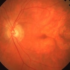 Central Areolar Choriocapillaris Atrophy
Mar 26 2019 by Gary R. Cook, MD, FACS
Left eye of a 64-year-old male with central (regional) areolar choroidal dystrophy showing fairly well circumscribed atrophy of the RPE and choriocapillaris in the macula; VA= 20/30
Imaging device: Topcon VT-50
Condition/keywords: choriocapillaris, hereditary choroidal atrophy, hereditary choroidal dystrophy
-
 Central Areolar Choriocapillaris Atrophy
Mar 26 2019 by Gary R. Cook, MD, FACS
Late-phase fluorescein angiogram image of the right eye of a 64-year-old white male with central areolar choriocapillaris atrophy showing late leakage from intact choriocapillaris around the perimeter of the disc and macular areas of choriocapillaris atrophy; VA= 20/50
Imaging device: Topcon VT-50
Condition/keywords: FA late phase, fluorescein angiogram (FA), hereditary choroidal atrophy, hereditary choroidal dystrophy
-
 Central Areolar Choriocapillaris Atrophy
Mar 26 2019 by Gary R. Cook, MD, FACS
Right eye of a 64-year-old male with central (or regional) atrophy of the RPE and choriocapillaris in the macula; VA= 20/30.
Imaging device: Topcon VT-50
Condition/keywords: choriocapillaris, hereditary choroidal atrophy, hereditary choroidal dystrophy
-
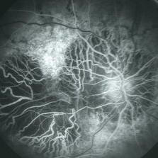
 Regional Choriocapillaris Atrophy
Mar 26 2019 by Gary R. Cook, MD, FACS
Late-phase (5 minutes) FA frame of the left eye of a 73-year-old white female with regional choriocapillaris atrophy showing light staining from intact choriocapillaris around the margins of the peripapillary and macular areas of RPE and choriocapillaris atrophy; VA= 20/100.
Imaging device: Topcon VT-50
Condition/keywords: atrophy, choriocapillaris, FA late phase, fluorescein angiogram (FA), hereditary choroidal atrophy, hereditary choroidal dystrophy
-
 Regional Choriocapillaris Atrophy
Mar 26 2019 by Gary R. Cook, MD, FACS
73-year-old white female with regional choriocapillaris atrophy OS; VA= 20/100.
Imaging device: Topcon VT-50
Condition/keywords: atrophy, choriocapillaris, hereditary choroidal atrophy, hereditary choroidal dystrophy
-
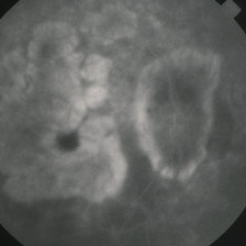
 Regional Choriocapillaris Atrophy
Mar 26 2019 by Gary R. Cook, MD, FACS
Mid-phase (laminar venous return phase) FA frame demonstrating thinning/loss of the RPE, choriocapillaris loss, and increased visibility of the larger choroidal vessels around the disc and in the macula from a 73-year-old white female with regional choriocapillaris atrophy; VA= 20/100.
Imaging device: Topcon VT-50
Condition/keywords: atrophy, choriocapillaris, fluorescein angiogram (FA), hereditary choroidal atrophy, hereditary choroidal dystrophy
-
 Regional Choriocapillaris Atrophy
Mar 26 2019 by Gary R. Cook, MD, FACS
73-year-old white female with regional choriocapillaris atrophy in the posterior pole of her right eye; VA= 20/100.
Imaging device: Topcon VT-50
Condition/keywords: atrophy, choriocapillaris, hereditary choroidal atrophy, hereditary choroidal dystrophy
 Loading…