Search results (113 results)
-
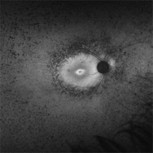
 Not All Stars Are in the Sky — Some Live in the Eyes of Those Learning to See in New Ways
Apr 21 2025 by rohan jain
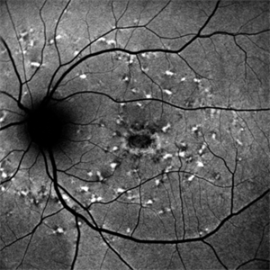
Stargardt disease
Photographer: Dr. ROHAN JAIN
Condition/keywords: fleck retinopathy, fundus autofluorescence (FAF), hereditary macular dystrophy
-
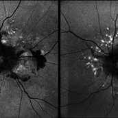
 Elmiron Toxicity
Mar 25 2025 by Toolie Winters
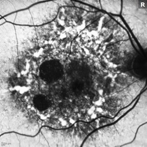
Fundus autofluorescence image of a 69-year-old woman with toxic maculopathy OU due to Elmiron usage. Patient stopped using Elmiron in the late 2010s after having been on it for 17 years. The patient has areas of outer retinal and RPE atrophy temporal to fovea that have expanded compared to photos from two years ago. At the time of this appointment, her VA OD was sc20/40-1+2 PH20/30 and VA OS was scCF @ 1 foot.
Photographer: Toolie Winters
Imaging device: Heidelberg Spectralis
Condition/keywords: Elmiron Toxicity, FAF, fundus autofluorescence (FAF), Heidelburg Spectralis, Pentosan Toxicity, Toxic Maculopathy
-
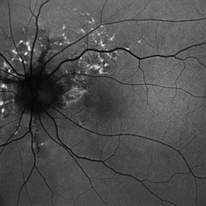
 FAF-G Circumscribed Choroidal Hemangioma
Mar 1 2025 by Vishal Agrawal, MD, FRCS,FACS,FASRS
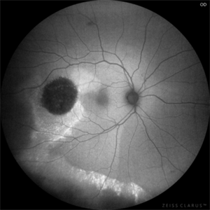
A 37-year-old male presented with decreased vision in the right eye. This is the fundus autofluorescence (FAF-G) of the right eye showing hypo auto fluorescent lesion with surrounding hyper auto fluorescence extending inferiorly corresponding to the fluid tract.
Photographer: Dr Ayushi Gupta
Imaging device: Clarus 700
Condition/keywords: Circumscribed Choroidal Hemangioma, fundus autofluorescence (FAF)
-
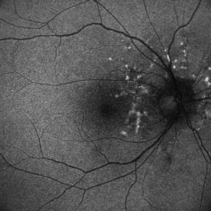
 Astrocytic Hamartoma
Feb 27 2025 by Daniel Davis, OCT-C
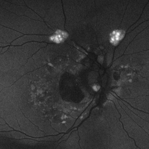
Fundus autofluorescence photo of 55-year-old female with astrocytic hamartoma in association with tuberous sclerosis. No treatment options available, benign. Other findings include; Posterior Vitreous Detachment, Vitreous Hemorrhage, Hereditary Retinal Dystrophy, Vitreous Opacities, Hypertensive Retinopathy.
Photographer: Daniel Davis, OCT-C
Imaging device: Optos California
Condition/keywords: astrocytic hamartoma, fundus autofluorescence (FAF)
-
 Retinitis Pigmentosa Bullseye Appearing Autofluorescence
Feb 4 2025 by Isaac Agranoff
Fundus Autofluorescence of a 14-year-old boy with suspected RP. ERG performed afterwards was almost flat. VA measured at 20/30 but with extensive constriction of confrontational visual fields. Currently awaiting genetic testing.
Photographer: Isaac Agranoff
Imaging device: Optos California
Condition/keywords: fundus autofluorescence (FAF), retinitis pigmentosa, RP
-
 Guardian Angel
Dec 11 2024 by Virginia Gebhart
48 year old female 3 months s/p brachytherapy for choroidal melanoma. Persistent subretinal and increased subfoveal fluid. Will observe for now, will consider Ozurdex if no improvement. BCVA 20/80
Photographer: Virginia Gebhart, Retina Consultants of Carolina
Imaging device: Optos California
Condition/keywords: brachytherapy, demarcation line, fundus autofluorescence (FAF), serous detachment, subretinal fluid
-
 Both Eyes Fundus Autofluorescence in Case of CNVM with Angioid Streaks
Nov 29 2024 by Anand Temkar
A 45 year old male came with chief complaint of blurring vision in right eyes since past 4 days. His vision is 6/12 in right eye and 6/9 in left eye. His vision was 14 mmHg in right eye and 16 mmHg in left eye. He was diagnosed with Angioid Streaks in both eyes about a year ago, then he developed choroidal neovascularization in his left eye 8 months ago, for which he received AntiVEGF injections x 3. Left eye is a stable eye now. Patient presented with right eye choroidal neovascularization in a case of Angioid Streaks on recent follow up. We have advised him right eye AntiVEGF injections x 3. In this image we can see fundus hypoautofluorescence in right eye due to hemorrhages and angioid streaks and in left eye fundus hypoautofluorescence is noted due to angioid streaks.
Photographer: Dr.Anand Temkar- Retina Foundation, Ahmedabad
Imaging device: Mirante
Condition/keywords: Angioid Streaks, choroidal neovascular membrane (CNVM), fundus autofluorescence (FAF)
-
 Fundus Flavimaculatus Fundus Autofluorescence Imaging
Sep 25 2024 by Keshavi Shah
FAF imaging of a 37 year old male patient with Stargardt's Disease of adult onset ( Fundus Flavimaculatus) presenting with dimunition of night vision and dyschromatopsia demonstrating areas of hypo-auto fluorescence (representing RPE/ Photo-receptor atrophy) and hyper-autofluorescence(representing excessive lipo-fuschin accumulation in the RPE cells) with peri-papillary sparing, typical of ABCA-4 related disorders.
Photographer: Simran
Imaging device: Optos Daytona
Condition/keywords: fundus autofluorescence (FAF), fundus flavimaculatus
-
 Fundus Flavimaculatus Fundus Autofluorescence Imaging
Sep 25 2024 by Keshavi Shah
FAF imaging of a 37 year old male patient with Stargardt's Disease of adult onset ( Fundus Flavimaculatus) presenting with dimunition of night vision and dyschromatopsia demonstrating areas of hypo-auto fluorescence (representing RPE/ Photo-receptor atrophy) and hyper-autofluorescence(representing excessive lipo-fuschin accumulation in the RPE cells) with peri-papillary sparing, typical of ABCA-4 related disorders.
Photographer: Simran
Imaging device: Nikon Optos Daytona
Condition/keywords: fundus autofluorescence (FAF), fundus flavimaculatus
-
 Fundus Autofluorescence Showing Angioid Streaks with Regressing CNV s/p AntiVEGF Injections (LE)
Sep 20 2024 by Anand Temkar
A 45 year old male came to our OPD with chief complaints of DOV in BE since 2 months and wavy vision in periphery. Patient was diagnosed with (BE) CNVM in a case of Angioid Streaks and has already received (BE) bevacizumab x 2.
Photographer: Dr.Anand Temkar- Retina Foundation, Ahmedabad
Imaging device: Mirante
Condition/keywords: Angioid Streaks, choroidal neovascularization (CNV), fundus autofluorescence (FAF)
-
 Fundus Autofluorescence Showing Angioid Streaks with Regressing CNV s/p AntiVEGF Injections (RE)
Sep 20 2024 by Anand Temkar
A 45 year old male came to our OPD with chief complaints of DOV in BE since 2 months and wavy vision in periphery. Patient was diagnosed with (BE) CNVM in a case of Angioid Streaks and has already received (BE) bevacizumab x 2.
Photographer: Dr.Anand Temkar- Retina Foundation, Ahmedabad
Imaging device: Mirante
Condition/keywords: Angioid Streaks, choroidal neovascularization (CNV), fundus autofluorescence (FAF)
-
 Angioid Streaks with Regressing CNV s/p AntiVEGF Injections (LE)
Sep 20 2024 by Anand Temkar
A 45 year old male came to our OPD with chief complaints of DOV in BE since 2 months and wavy vision in periphery. Patient was diagnosed with (BE) CNVM in a case of Angioid Streaks and has already received (BE) bevacizumab x 2.
Photographer: Dr.Anand Temkar- Retina Foundation, Ahmedabad
Imaging device: Mirante
Condition/keywords: Angioid Streaks, choroidal neovascularization (CNV), fundus autofluorescence (FAF)
-
 Angioid Streaks with Regressing CNV s/p AntiVEGF Injections (RE)
Sep 20 2024 by Anand Temkar
A 45 year old male came to our OPD with chief complaints of DOV in BE since 2 months and wavy vision in periphery. Patient was diagnosed with (BE) CNVM in a case of Angioid Streaks and has already received (BE) bevacizumab x 2.
Photographer: Dr.Anand Temkar- Retina Foundation, Ahmedabad
Imaging device: Mirante
Condition/keywords: Angioid Streaks, choroidal neovascularization (CNV), fundus autofluorescence (FAF)
-
 Pericentral Retinitis Pigmentosa
Sep 6 2024 by Mauricio Bayram-Suverza, MD
A 65-year-old male patient reports experiencing bilateral blind spots that have gradually intensified over time. Genetic testing was unrevealing. The fundus autofluorescence image shows a hypoautofluorescent ring in the posterior pole, especially nasal to the nerve and along arcades.
Photographer: Mauricio Bayram-Suverza, Casey Eye Institute, OHSU.
Imaging device: Optos California
Condition/keywords: fundus autofluorescence (FAF), inherited retinal disease, nyctalopia, retinal dystrophy, retinitis pigmentosa
-
 Stargardt Disease
Aug 27 2024 by Korey Starkey
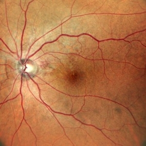
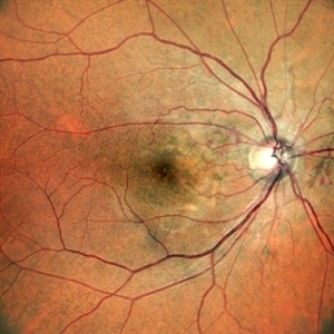
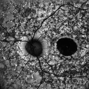
Ultra wide-field fundus photograph and fundus autofluorescence of a 49-year-old male. Initial visit imaging.
Photographer: Korey Starkey
Imaging device: Optos
Condition/keywords: fundus autofluorescence (FAF), fundus photograph, Optos, Stargardt disease, ultra-wide field imaging
-
 B-FAF in Stargardt's Disease
Jul 4 2024 by Tejaswita Verma
Blue fundus autofluorescence showing hypoautofluorescence picture of a 28 year old male with 6/60 vision in BE in a case of Stargardt's disease.
Photographer: DR. TEJASWITA VERMA
Imaging device: MIRANTE
Condition/keywords: fundus autofluorescence (FAF), hereditary macular dystrophy, Stargardt disease
-
 FAF of Barricade Laser on Choroidal Osteoma
Jun 12 2024 by Virginia Gebhart
20 year old female with stable choroidal osteoma s/p PDT x 3 and focal laser x 2. No obvious progression on last exam, vision 20/30. Monitoring closely.
Photographer: Virginia Gebhart
Imaging device: Topcon 50 DX
Condition/keywords: autofluorescence imaging, barrier laser, choroidal osteoma, focal laser, fundus autofluorescence (FAF)
-
 Revealing the Invisible
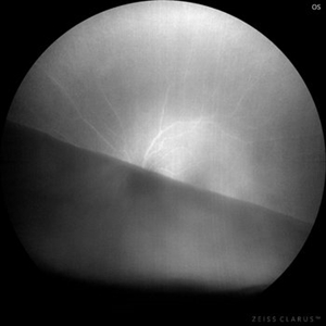
Apr 25 2024 by Ricardo Leitão Guerra
In this remarkable wide-field autofluorescence image, we observe a patient with a giant retinal tear that has precipitated both retinal detachment and subsequent folding upon itself. Notably, the image distinctly maps the original anatomical configuration of the blood vessels within the affected retina.
Photographer: Ricardo Leitão Guerra, Leitão Guerra - Oftalmologia.
Imaging device: Clarus 700 - Zeiss Medical technology.
Condition/keywords: fundus autofluorescence (FAF), GIANT RETINAL TEAR
-
 Stargardt's Disease
Apr 20 2024 by Tejaswita Verma
Fundus autofluorescence image of the left eye of a 39 year old male showing hypoautofluorescence in a case of Stargardt's disease.
Photographer: DR. TEJASWITA VERMA
Imaging device: MIRANTE
Condition/keywords: fundus autofluorescence (FAF), hereditary macular dystrophy, hypoautofluorescence, Stargardt disease
-
 Stargardt's Disease
Apr 20 2024 by Tejaswita Verma
Fundus autofluorescence image of the right eye of a 39 year old male showing hypoautofluorescence in a case of Stargardt's disease.
Photographer: DR. TEJASWITA VERMA
Imaging device: MIRANTE
Condition/keywords: fundus autofluorescence (FAF), hereditary macular dystrophy, hypoautofluorescence, Stargardt disease
-
 Choroidal Metastasis
Apr 11 2024 by Corey Grant
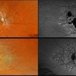
Ultra-Widefield fundus photography and fundus autofluorescence images of a 61 year old female with Choroidal Metastasis affecting both eyes. Patient presented with blurred vision and flashes for a few weeks. Patient visual acuity was cc20/100 PH20/60 in the right eye and cc20/200 in the left eye. Patient admits to history of smoking for many years bit no known history of cancer prior to the visit. Physician recommended going to the ER for full body PET CT and stated that the first line of treatment is usually systemic chemo therapy. Patient will be reassessed in one month.
Photographer: Corey Grant
Imaging device: OPTOS CALIFORNIA RGB
Condition/keywords: cancer, choroidal metastasis, fundus autofluorescence (FAF), fundus photography, hyperautofluorescence, hypoautofluorescence, Optos, OPTOS CALIFORNIA RGB, Retina, ULTRA WIDE FIELD
-
 Stargardt Disease
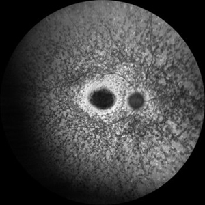
Apr 8 2024 by T. P . VIGNESH, MBBS,MS
Fundus autofluorescence image of the left eye revealing foveal hypoautofluorescence and multiple hypoautofluorescent specks in the background radiating from posterior pole towards periphery .
Photographer: Bharathi
Imaging device: ZEISS CLARUS
Condition/keywords: fundus autofluorescence (FAF), Stargardts Disease
-
 Stargardt Disease
Apr 8 2024 by T. P . VIGNESH, MBBS,MS
Fundus autofluorescence image of the right eye revealing foveal hypoautofluorescence and multiple hypoautofluorescent specks in the background radiating from posterior pole towards periphery.
Photographer: Bharathi
Imaging device: ZEISS CLARUS
Condition/keywords: fundus autofluorescence (FAF), Stargardt disease
-
 Central Serous Retinopathy
Mar 19 2024 by Corey Grant
Ultra Wide-Field Fundus Autofluorescence Imaging of a 37 year old female with Central Serous Retinopathy affecting her right eye. Patient Visual Acuity was 20/20 in both eyes. Patient reported black spots in her vision onset three years ago, with associating flashes of light. Patient reports history of cortisone back injections a few years ago and denies Flonase use. The physician stated that there is hyperautofluorescence in the area of gutter of Sub-Retinal Fluid which likely happened from CSR.
Photographer: Corey Grant, OSC
Imaging device: OPTOS CALIFORNIA RGB
Condition/keywords: Central Serous Chorioretinopathy (CSR), central serous retinopathy (CSR), fundus autofluorescence (FAF), Guttering, hyperautofluorescence, inferior retina, OPTOS, Retina, Right Eye, subretinal fluid, ULTRA WIDE FIELD
-
 Multimodal Imaging for Differentiating Unilateral Pseudo Optic Disc Swelling(Buried Drusen) From True Optic Disc Swelling
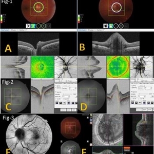
Feb 7 2024 by Fawwaz F Al Mamoori, MD, Medical Retina Consultant
27-year-old male, medically free, presented with left unilateral optic disc swelling. BCVA=1.0(OU), color vision, and contrast sensitivity were normal (OU)with no RAPD in the left eye. Swept Source OCT: showed elevated left optic disc with hyporeflective mass (Fig-1 B). Enface OCT: Showed left peripapillary multiple ovoid mass lesions(drusen) (Fig-2 d, Fig3 F). FAF: of the left eye showed superonasal hyper autofluorescent drusenoid lesions)(Fig3 E). Orbital MRI with contrast was requested to exclude any compressive lesions like tumors(menigioma)or inflammatory lesions like granuloma(sarcoid granuloma). orbital MRI result was normal.
Photographer: Hana.S.Owais
Imaging device: TRITON(TOPCON,Swept Source OCT)
Condition/keywords: fundus autofluorescence (FAF), multimodal imaging, OCT EN FACE, optic disc drusen, optic disc edema, swept source
 Loading…