Search results (6 results)
-
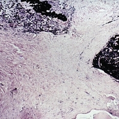
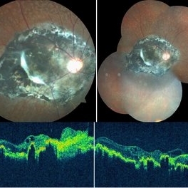 Chorioretinitis Sclopetaria
May 4 2021 by Priya Rasipuram Chandrasekaran, MBBS, DO, DNB, FRCS
This fundus photo and montage shows pigmentary changes with fibroglial proliferation of the disc and macula in a 36-year-old male following injury with an iron chain. This is usually following a high velocity non-penetrating missile or blast injury categorized as coup injury and can be both direct or indirect. The layers affected are the highly inelastic Bruch’s membrane with choriocapillaris and retinal pigment epithelium in contrast to the highly elastic retina and sclera. The high impact injury causes full thickness defect in the retina, Bruch’s membrane and choroid leading to retraction of the retina and choroid, leaving the intact bare sclera behind. Pathology included defects in the Bruch’s membrane and choroid, and extensive photoreceptor loss with hyperplasia of retinal pigment epithelium. Over the weeks, loose fibrous tissue gets replaced by dense connective tissue leading to scarring between retina and choroid as seen in our patient. The background shows fundus albipunctatus.
Condition/keywords: chorioretinitis sclopetaria
-
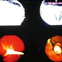 Slide 8-20
Mar 4 2019 by Lancaster Course in Ophthalmology
Examples of fibrous tissue proliferation in the vitreous at sites of penetration of globe. Fibrous encasement of a retained intraocular foreign body is illustrated in the lower right view.
Condition/keywords: fibrous tissue proliferation
-
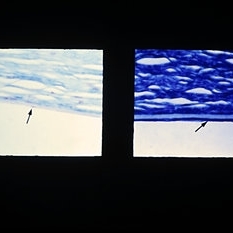 Slide 8-17
Mar 4 2019 by Lancaster Course in Ophthalmology
Right: Aphakic bullous keratopathy with vitreous touch. The endothelium is markedly flattened with extreme irregular spacing of nuclei. One flattened endothelial-cell nucleus is present (arrow) (E.P. No. 39964) Left: Aphakic bullous keratopathy with vitreous touch, partial endothelial atrophy, and retrocorneal fibrous tissue proliferation (arrow). (E.P. No. 39234)
Condition/keywords: atrophy, keratopathy, retrocorneal fibrous tissue proliferation, vitreous touch
-
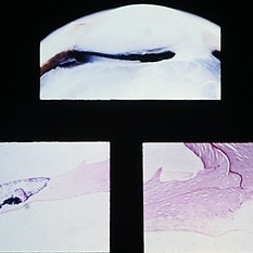 Slide 8-16
Mar 4 2019 by Lancaster Course in Ophthalmology
Updrawn pupil following cataract extraction. Vitreous is incarcerated in the cataract scar above. The inferior iris leaf is adherent to fibrous tissue that has proliferated along the tract of the anterior vitreous. Contraction of this fibrosed anterior vitreous has pulled the attached inferior iris leaf toward the wound and has resulted in an updrawn pupil. (E.P. No. 31006)
Condition/keywords: cataract, cataract extraction, fibrous tissue, iris leaf, updrawn pupil, vitreous
-
 Slide 7-103
Feb 25 2019 by Lancaster Course in Ophthalmology
Fibrous downgrowth. Fibrous tissue extends through the iridectomy site into the posterior chamber.
Condition/keywords: Fibrous downgrowth, peripheral iridotomy
-
 PHPV
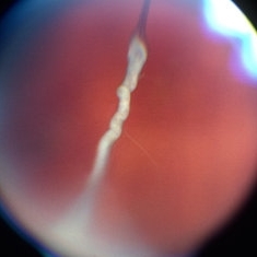
PHPV
May 2 2013 by Henry J. Kaplan, MD
The same patient; hyaloid artery has changed to fibrous tissue anteriorly; #3.
Condition/keywords: hyaloid artery, persistent fetal vasculature (PFV), persistent hyperplastic primary vitreous (PHPV)
 Loading…