Search results (121 results)
-
 Diabetic Macular Edema
Jul 3 2025 by Gustavo Uriel Fonseca Aguirre
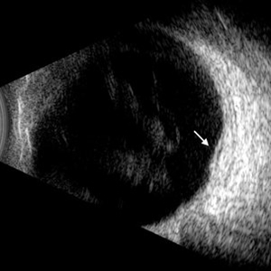
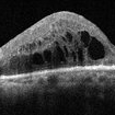
This B-mode longitudinal ultrasound scan demonstrates diabetic macular edema with mild subretinal fluid accumulation, appearing as a subtle hypoechoic space beneath the neurosensory retina. The macular region shows retinal thickening and heterogeneous medium reflectivity, consistent with active exudative changes (arrow). No vitreomacular traction is observed.
Photographer: Gustavo U. Fonseca Aguirre, Hospital Conde de Valenciana, Ciudad de México
Condition/keywords: diabetic macular edema
-
 Diabetic Macular Edema
Apr 28 2025 by Gustavo Uriel Fonseca Aguirre
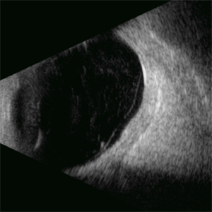
This B-mode longitudinal ultrasound scan demonstrates irregular macular thickening with homogeneous medium-to-high internal reflectivity, consistent with diabetic macular edema. The lesion shows poorly defined borders and absence of cystic spaces or subretinal fluid on dynamic evaluation.
Photographer: Gustavo U. Fonseca Aguirre, Hospital Conde de Valenciana, Ciudad de México
Condition/keywords: diabetic macular edema
-
 Ozurdex Implant
Mar 12 2025 by Virginia Gebhart
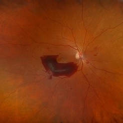
65 year old female 4 weeks s/p intravitreal Ozurdex implant for diabetic macular edema. Significant improvement of edema and SRF
Photographer: Virginia Gebhart, Retina Consultants of Carolina
Imaging device: Optos California
Condition/keywords: diabetic macular edema, DME, intravitreal implant, intravitreal injection, ozurdex, Ozurdex implant
-
 Diabetic Macular Edema
Feb 12 2025 by Kimberly Wakester
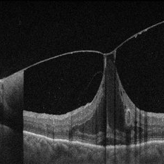
Horizontal OCT scan of a 63-year-old woman with diabetic macular edema in the right eye. When reviewing the scan, one of the intraretinal cyst (IRC) appears heart shaped. A fun scan to see just a few day's before Valentine's day.
Photographer: Kimberly Wakester, COA
Imaging device: Heidelberg
Condition/keywords: diabetic macular edema, intraretinal cyst
-
 Vitreomacular Adhesion Showcasing a Microaneurysm and a Subhyaloid Hemorrhage
Jan 3 2025 by Drew Mitchell
A vertical OCT 1 line raster scan positioned slightly inferomacula to document the subhyaloid hemorrhage. Hyper reflective Oval indicating Microaneurysm.
Photographer: Drew Mitchell, OCT-C
Imaging device: Zeiss Cirrus 5000
Condition/keywords: diabetic macular edema, microaneurysms, retinal microaneurysms, subhyaloid hemorrhage, subretinal fluid, vitreomacular adhesion, vitreomacular traction (VMT)
-
 Pre-Retinal Hemorrhage
Aug 22 2024 by Virginia Gebhart
51 year old female with moderate proliferative diabetic retinopathy, DME, as well as pre-retinal hemorrhage and likely NVE. Pt given Avastin in office and will return for PRP.
Photographer: Virginia Gebhart
Imaging device: Optos California
Condition/keywords: diabetic macular edema, macular edema, PDR with NVE (periphery), pre-retinal hemorrhage, proliferative diabetic retinopathy (PDR)
-
 Floating Ozurdex Implant
May 20 2024 by Tejaswita Verma
Fundus photograph of the left eye of a 73 year old female with ozurdex implant floating in the vitreous in a diabetic lasered patient.
Photographer: DR. TEJASWITA VERMA
Imaging device: MIRANTE
Condition/keywords: diabetic macular edema, laser photocoagulation, Ozurdex implant
-
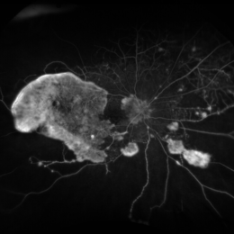
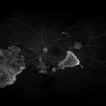
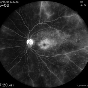 Angiographic Diabetic Macular Edema in a Case of Proliferative Diabetic Retinopathy
Apr 9 2024 by Akansha Sharma
Fundus fluorescein angiographic image of 62 year old male demonstrating angiographic diabetic macular edema in a case of proliferative diabetic retinopathy.
Photographer: Dr. Akansha Sharma, Bharati Eye Hospital
Condition/keywords: clinically significant macular edema (CSME), diabetic blindness, diabetic macular edema, proliferative diabetic retinopathy (PDR)
-
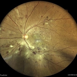
 The Starry Sky
Mar 12 2024 by MEENAL SONI
57 year old female with DOV in BE, known diabetic. OU Multiple microaneurysms and haemorrhages at macula with diabetic macular oedema.
Photographer: Dr. Meenal Soni, Fellow VR, ASG eye Hospital Jodhpur
Imaging device: ZEISS Visucam 400
Condition/keywords: diabetic macular edema, Diabetic Retinopathy
-
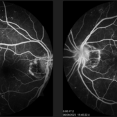
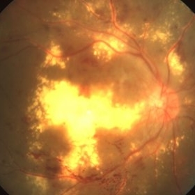
 Serous Retinal Detachment in Advanced Proliferative Diabetic Retinopathy
Feb 15 2024 by Annaka Gooding
Ultra-Wide fundus photograph of a 29 year old female with a Serous Retinal Detachment in Advanced PDR. Patient present to clinic with LP vision following PPV and fill in PRP. Physician recommended oral prednisone treatment and to reassess at their following visit.
Photographer: Annaka Gooding, CPO
Imaging device: Optos California RGB
Condition/keywords: Diabetes, diabetic macular edema, fundus photography, OPTOS CALIFORNIA, pan-retinal photocoagulation (PRP), pars plana vitrectomy (PPV), proliferative diabetic retinopathy (PDR), serous retinal detachment, ultra-wide field imaging
-
 Diabetic Macular Edema
Feb 7 2024 by Virginia Gebhart
FA of 70 year old male with diabetic macular edema. FA shows early hyper-fluorescence with late leakage and capillary dropout in the temporal macula. Focal laser performed.
Photographer: Virginia Gebhart
Imaging device: Optos California
Condition/keywords: capillary dropouts, macular edema
-
 Bullseye Maculopathy
Jan 22 2024 by Kali Jend
Optical coherence tomography of a 73-year-old female with Bullseye Macular Changes affecting her left eye. Patient reports having a family history of this condition and denies prior Plaquenil or Elmiron use. Compared to previous imaging, the patient's condition progressed in the left eye from 2020 to 2023. Patient has a history of fluctuating Diabetic Macular Edema and a current Epiretinal Membrane as well. Patient's vision was Ncc20/60 at the time the image was taken.
Photographer: Kali Jend
Imaging device: Heidelberg Spectralis
Condition/keywords: bullseye maculopathy, epiretinal membrane (ERM), heidelberg spectralis, left eye, macular pucker, OCT, optical coherence tomography (OCT)
-
 Severe NPDR
Oct 24 2023 by Virginia Gebhart
Fluorescein angiogram of left eye in 60-year-old male with severe non-proliferative diabetic retinopathy with extensive macular edema. Most recent A1c is 11. Vision 20/400. Injection of Eylea given
Photographer: Virginia Gebhart
Imaging device: Topcon
Condition/keywords: diabetic macular edema, Diabetic Retinopathy
-
 Ozurdex implant for diabetic macular oedema
Sep 6 2023 by PRATIK SHENOY, MBBS, DNB, FVRS
A 53-year-old female presented with diabetic macular oedema. She had undergone pan-retinal photocoagulation for proliferative diabetic retinopathy previously. She was injected with an intravitreal ozurdex implant for the same.
Photographer: Gaurav Kamble, Isha Netralaya
Imaging device: Optos
Condition/keywords: diabetic macular edema, Optos, ozurdex, pan-retinal photocoagulation (PRP), proliferative diabetic retinopathy (PDR)
-
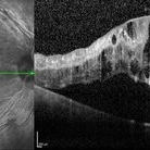
 Spent-force
Jun 27 2023 by Maneesh M Bapaye, MD, MBA
Composite OCT image of left eye of a 53 years old male diabetic patient with recuuent spongy diabetic macular edema. Structural image depicts presence of ozurdex temporal to fovea. B-Scan image shows localized action of ozurdex in retinal tissue underlying it while cystoid changes are seen nasal to foveal center
Photographer: Maneesh Bapaye MD
Condition/keywords: diabetic macular edema, Ozurdex implant
-
 PROLIFERATIVE DIABETIC RETINOPATHY
Jun 6 2023 by Akansha Sharma
COLOUR FUNDUS PHOTOGRAPH OF A 42 YEAR OLD DIABETIC MALE WITH PROLIFERATIVE DIABETIC RETINOPATHY AND MACULAR EDEMA
Photographer: Dr. Denish Patel, Dr. Akansha Sharma, Dr. Urmil Shah, Bharati Eye Hospital, Ahmedabad, Gujarat
Condition/keywords: diabetes, diabetic macular edema, macular edema, PDR, proliferative diabetic retinopathy (PDR)
-
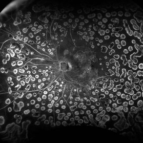
 Combined Tractional and Rhegmatogenous Retinal Detachment
Jan 30 2023 by Olivia Rainey
Ultra-widefield fluorescein angiography of a combined tractional and rhegmatogenous retinal detachment repair affecting the left eye. The retina is attached following silicone oil placement during most recent surgery. The patient was seeing CF at the time the image was taken.
Photographer: Olivia Rainey, OCT-C, COA
Imaging device: Optos California
Condition/keywords: diabetes, diabetic macular edema, diabetic retinopathy, hyperfluorescence, right eye, scleral buckle, silicone oil, tractional retinal detachment, ultra-wide field imaging, ultra-widefield image
-
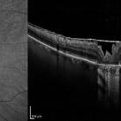
 Proliferative diabetic retinopathy with diabetic macular edema
Oct 21 2022 by T. P . VIGNESH, MBBS,MS
SD-OCT of a 50 year man with PDR and massive macular edema with sub foveal detachment and hard exudates, Foveal Thickness -1418 microns.
Photographer: Shivanath
Imaging device: Heidelberg Spectralis
Condition/keywords: proliferative diabetic retinopathy (PDR)
-
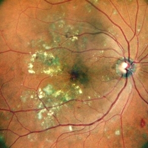 CIRCINATE RETINOPATHY
Oct 19 2022 by Akansha Sharma
COLOUR FUNDUS PHOTOGRAPH OF A 51 YEAR OLD MALE WITH DIABETIC MACULOPATHY
Photographer: Dr. Akansha Sharma-Retina Foundation, Ahmedabad
Condition/keywords: circinate retinopathy, diabetic macular edema, diabetic maculopathy
-
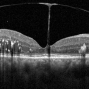 Vitreomacular traction
Jun 23 2022 by T. P . VIGNESH, MBBS,MS
SD-OCT of RE reveals Vitreomacular traction resembling bow and arrow and diabetic macular edema with intraretinal hard exudates in a 60 year old female patient with Moderate NPDR .
Imaging device: Heidelberg Spectralis
Condition/keywords: vitreomacular traction (VMT)
-
 Proliferative diabetic retinopathy with dense macular hard exudates
Mar 30 2022 by T. P . VIGNESH, MBBS,MS
Fundus photo of a 60 year old male patient with Proliferative diabetic retinopathy and dense macular hard exudates .
Photographer: Bharathi Singaravel
Imaging device: Zeiss FF450plus IR
Condition/keywords: diabetic macular edema, Diabetic Retinopathy
-
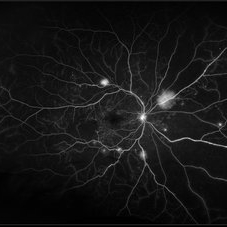
 Severe Diabetic Retinopathy, Nonperfusion and NVE
Jun 18 2021 by Kristen Wagner
Optos angiogram of severe diabetic retinopathy, nonperfusion and NVE.
Photographer: Kristen Wagner, COT Tennessee Retina Nashville TN
Imaging device: Optos
Condition/keywords: diabetic macular edema, neovascularization elsewhere (NVE), nonperfusion diabetic retinopathy, proliferative diabetic retinopathy (PDR)
-
 Severe Diabetic Retinopathy and NVE with Macular Hole
Jun 18 2021 by Kristen Wagner
Fundus photo of severe PDR with NVE and nonperfusion. The patient also has a macular hole.
Photographer: Kristen Wagner, COT Tennessee Retina Nashville TN
Imaging device: Optos
Condition/keywords: diabetic macular edema, macular hole, neovascularization elsewhere (NVE), proliferative diabetic retinopathy (PDR)
-
 Diabetic Macular Edema
Mar 14 2021 by Marco Antonio Sauza
Diabetic Macular Edema
Photographer: Marco Sauza
Imaging device: Heidelberg
Condition/keywords: diabetic macular edema
-
 Proliferative Diabetic Retinopathy
Jan 29 2021 by Olivia Rainey
Ultra-widefield fluorescein angiogram of a 65-year-old male with proliferative diabetic retinopathy affecting his right eye. The patient's diabetic retinopathy has progressed significantly since he was last seen in 2014. It was recommended to begin antiVEGF to control DME followed by laser treatment OU.
Photographer: Olivia Rainey, OCT-C, COA
Imaging device: Optos California
Condition/keywords: anti-VEGF, diabetes, diabetic macular edema, neovascularization (NV), neovascularization elsewhere (NVE), non-perfusion, Optos, proliferative diabetic retinopathy (PDR), ultra-wide field imaging
 Loading…