Search results (544 results)
-
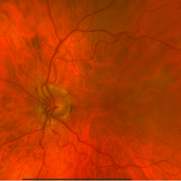
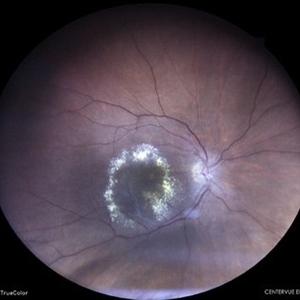
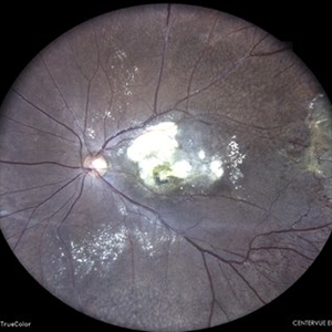
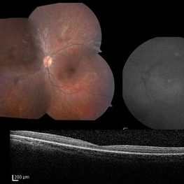 LCA Type 2
Apr 10 2025 by Joshua Friedman
LCA Type 2 (RPE65) showing characteristic hypoautofluorescence and retinal thinning. 8F with best corrected visual acuity of 20/400 (OD) and 20/150 (OS). Small white intraretinal spots and RPE mottling are visible on color fundus photography. Blue light autofluorescence reveals near-complete loss of signal, while OCT demonstrates widespread outer retinal thinning.
Photographer: Stephen Tsang, MD, PhD
Condition/keywords: Leber Congenital Amaurosis
-
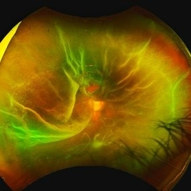
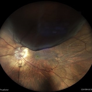
 MacroAneurysm - 1 Day After Rupture
Mar 31 2025 by Max Whitmeyer
Fundus photograph of a macroaneurysm progression prior to and following rupture.
Photographer: Natasa Stankovich, Edward Hines Jr. VA Hospital
Imaging device: Zeiss Clarus 500
Condition/keywords: color fundus photograph, macroaneurysm
-
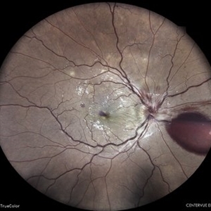
 MacroAneurysm - 1 Week Before Rupture
Mar 31 2025 by Max Whitmeyer
Fundus photograph of a macroaneurysm progression prior to and following rupture.
Photographer: Natasa Stankovich, Edward Hines Jr. VA Hospital
Imaging device: Zeiss Clarus 500
Condition/keywords: color fundus photograph, macroaneurysm
-
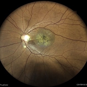
 MacroAneurysm - 2 Months Before Rupture
Mar 31 2025 by Max Whitmeyer
Fundus photograph of a macroaneurysm progression prior to and following rupture.
Photographer: Natasa Stankovich, Edward Hines Jr. VA Hospital
Imaging device: Zeiss Clarus 500
Condition/keywords: color fundus photograph, macroaneurysm
-
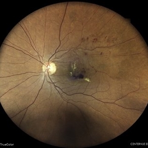
 MacroAneurysm - 3 Months Before Rupture
Mar 31 2025 by Max Whitmeyer
Fundus photograph of a macroaneurysm progression prior to and following rupture.
Photographer: Natasa Stankovich, Edward Hines Jr. VA Hospital
Imaging device: Zeiss Clarus 500
Condition/keywords: color fundus photograph, macroaneurysm
-
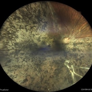
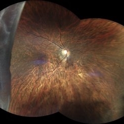
 Rhegmatogenous Retinal Detachment
Mar 24 2025 by DR APOORVA JADHAV, MBBS , DNB
This is a color fundus photograph showing rhegmatogenous retinal detachment with posterior pole retinal tear with macula off.
Condition/keywords: retinal detachment of the macula
-
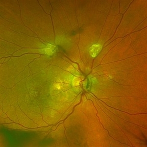 Astrocytic Hamartoma
Feb 27 2025 by Daniel Davis, OCT-C
Color fundus photo of 55-year-old female with Astrocytic Hamartoma in association with tuberous sclerosis. No treatment options available, benign. Other findings include; Posterior Vitreous Detachment, Vitreous Hemorrhage, Hereditary Retinal Dystrophy, Vitreous Opacities, Hypertensive Retinopathy.
Photographer: Daniel Davis, OCT-C
Imaging device: Optos California
Condition/keywords: color fundus photograph
-
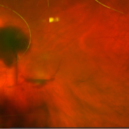
 Von Hippel-Lindau Syndrome
Jan 7 2025 by Jordyn Beckman
Fundus photograph of an 37 year old female presents with reddish vascular lesion with feeder vessels for possible Von Hippel-Lindau Syndrome.
Photographer: Jordyn Beckman
Imaging device: California Optos
Condition/keywords: color fundus photograph, feeder vessel, genetic disorder, pre-cryotherapy
-
 Tractional Retinal Detachment in a Case of Proliferative Diabetic Retinopathy
Aug 6 2024 by Akansha Sharma
Color fundus photograph of a 60 year old female with tractional retinal detachment in a case of proliferative diabetic retinopathy.
Photographer: Dr. Akansha Sharma, Bharati Eye Hospital
Condition/keywords: fibrovascular proliferation, PDR, Proliferative Diabetic retinopathy, tractional retinal detachment, TRD
-
 Spontaneously Settled Retinal Detachment
Aug 6 2024 by Akansha Sharma
Color fundus photograph of a 36 year old female with spontaneously settled retinal detachment.
Photographer: Dr. Akansha Sharma, Bharati Eye Hospital
Condition/keywords: OLD SETTLED RETINAL DETACHMENT, SPONTANEOUSLY SETTLED RETINAL DETACHMENT
-
 Retinal Detachment
Aug 6 2024 by Akansha Sharma
Color fundus photograph of a 36 year old male with retinal detachment.
Photographer: Dr. Akansha Sharma, Bharati Eye Hospital
Condition/keywords: RD, Retinal Detachment
-
 Venous Stasis Retinopathy
Aug 6 2024 by Akansha Sharma
Color fundus photograph of a 18 year old male with venous stasis retinopathy.
Photographer: Dr. Akansha Sharma, Bharati Eye Hospital
Condition/keywords: arterial occlusion, venous stasis retinopathy
-
 Circinate Retinopathy in a Case of Old Vein Occlusion
Aug 6 2024 by Akansha Sharma
Color fundus photograph of a 65 year old female with circinate retinopathy in a case of old vein occlusion.
Photographer: Dr. Akansha Sharma, Bharati Eye Hospital
Condition/keywords: circinate retinopathy, Vein Occlusion
-
 Coats' Disease
Aug 6 2024 by Akansha Sharma
Color fundus photograph of a 15 year old male with coats' disease.
Photographer: Dr. Akansha Sharma, Bharati Eye Hospital
Condition/keywords: Coats' disease
-
 Retinal Detachment With Choroidal Detachment
Aug 6 2024 by Akansha Sharma
Color fundus photograph of a 49 year old male with retinal detachment and choroidal detachment.
Photographer: Dr. Akansha Sharma, Bharati Eye Hospital
Condition/keywords: CD, choroidal detachment, RD, Retinal Detachment
-
 Retinal Detachment
Aug 6 2024 by Akansha Sharma
Color fundus photograph of a 39 year old male with retinal detachment status post trauma.
Photographer: Dr. Akansha Sharma, Bharati Eye Hospital
Condition/keywords: RD, Retinal Detachment
-
 A Fleet of Boat-Shaped Hemorrhages
Aug 1 2024 by James P Dossett, MD
Pseudocolor fundus photograph of the left eye of a 54-year-old diabetic man presenting with bilateral vision loss. Examination revealed 20/200 vision OS with extensive preretinal and vitreous hemorrhage, marked diffuse neovascularization, macular edema and hard exudates.
Photographer: Beth Smith, West Virginia University Eye Institute
Condition/keywords: proliferative diabetic retinopathy (PDR)
-
 Retinal Detachment
Jun 27 2024 by Akansha Sharma
Color fundus photograph of a 65 year old male with retinal detachment with horse shoe tear at 12 o'clock.
Photographer: Dr. Akansha Sharma, Bharati Eye Hospital
Condition/keywords: RD, Retinal Detachment
-
 Venous Stasis Retinopathy
Jun 27 2024 by Akansha Sharma
Color fundus photograph of a 31 year old male patient with venous stasis retinopathy.
Photographer: Dr. Akansha Sharma, Bharati Eye Hospital
Condition/keywords: cystoid macular edema (CME), Disc Edema, SHH, Sub hyaloid haemorrhage
-
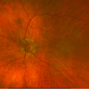
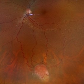
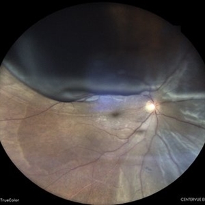
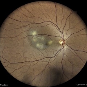
 Optic Disc Pit With Macular Scar
Jun 24 2024 by Akansha Sharma
Color fundus photograph of a 42 year old male with optic disc pit with macular scar.
Photographer: Dr. Akansha Sharma, Bharati Eye Hospital
Condition/keywords: macular scar, Optic disc pit
-
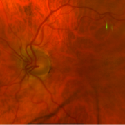
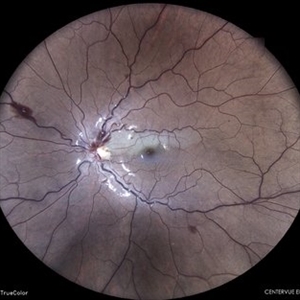
 Old Vein Occlusion With Macular Edema
Jun 24 2024 by Akansha Sharma
Color fundus photograph of a hypertensive 53 year old female with vein occlusion with macular edema.
Photographer: Dr. Akansha Sharma, Bharati Eye Hospital
Condition/keywords: macular edema, Vein Occlusion
-
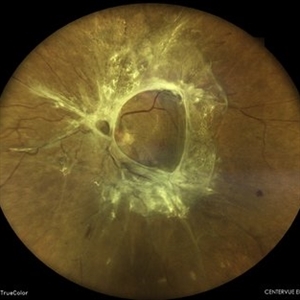
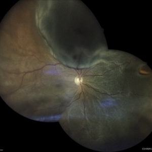
 Giant Retinal Tear
Jun 24 2024 by Akansha Sharma
Color fundus photograph of a 30 year old female with a giant retinal tear.
Photographer: Dr. Akansha Sharma, Bharati Eye Hospital
Condition/keywords: GIANT RETINAL TEAR, GRT
-
 Retinal Detachment
Jun 24 2024 by Akansha Sharma
Color fundus photograph of a 56 year old male with retinal detachment with visible horse shoe tear.
Photographer: Dr. Akansha Sharma, Bharati Eye Hospital
Condition/keywords: RD, Retinal Detachment
-
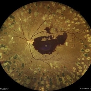 Subhyaloid Hemorrhage in a Case of Lasered Proliferative Diabetic Retinopathy
Jun 24 2024 by Akansha Sharma
Color fundus photograph of a 47 year old female with subhyaloid hemorrhage in a case of lasered proliferative diabetic retinopathy.
Photographer: Dr. Akansha Sharma, Bharati Eye Hospital
Condition/keywords: PDR, proliferative diabetic retinopathy (PDR), SHH, Sub hyaloid haemorrhage
-
 TB Choroiditis
Jun 17 2024 by Akansha Sharma
Color fundus photograph of a 40 year old male with TB choroiditis.
Photographer: Dr. Akansha Sharma, Bharati Eye Hospital
Condition/keywords: TB CHOROIDITIS
 Loading…