Search results (86 results)
-
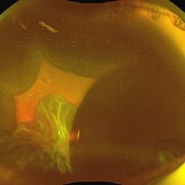
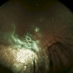
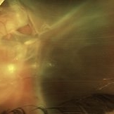
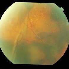
 Nail IOFB 2
Dec 30 2025 by Jared B Macher, MD
Zone 2 globe injury with nail IOFB. After removal patient was found to have a 3 by 6mm wound associated with a vitreous and suprachoroidal hemorrhage.
Photographer: Jared Macher, MD, University of Minnesota
Condition/keywords: IOFB, Penetrating trauma
-
 Kissing Choroidals
Dec 18 2025 by Talhah - Zubair, MD
72 year old woman developed suprachoroidal hemorrhage during Yamane scleral intraocular lens fixation. At clinic follow up they were found to be appositional. Suprachoroidal tissue plasminogen activator was injected in clinic and an emergent choroidal cutdown drainage was performed the following day with subsequent resolution of apposition. Appositional choroidals were managed urgently to avoid retinal adhesion/membrane formation.
Condition/keywords: appositional choroidals, kissing choroidals, suprachoroidal hemorrhage
-
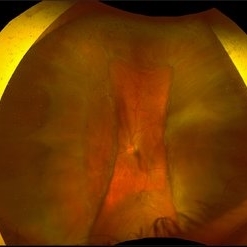
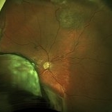
 Suprachoroidal Hemorrhage
Aug 4 2025 by Anjana Mirajkar, MS Ophthalmology
A fundus photograph of a 56 year old female with a 360 degree suprachoroidal hemorrhage with a 360 degree crumpled retina during cataract surgery.
Photographer: Dr. Anjana Mirajkar- HV Desai eye hospital ,Pune
Imaging device: optos
Condition/keywords: giant retinal tear, suprachoroidal hemorrhage
-
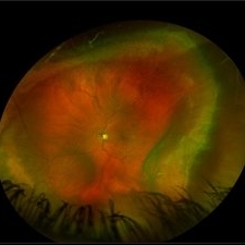
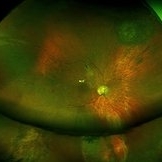
 Suprachoroidal Hemorrhage
Dec 3 2024 by Dibya Prabha
Colour Fundus photograph of 62 Year old female patient with Suprachoroidal hemorrhage post trauma
Photographer: Dibya Prabha, LV Prasad eye Institute, Hyderabad
Condition/keywords: suprachoroidal hemorrhage
-
 PEHCR
PEHCR
Jan 4 2024 by Virginia Gebhart
86 year old male with partially oxidized choroidal hemorrhage and CME. Previous FA shows blocking defect temporally, most likely a choroidal hemorrhage with SRH and late leakage. Continued improvement with 8 week intervals of Eylea. VA 20/60 Previous RD repair with scleral buckle and cryo in 1980's
Photographer: Virginia Gebhart
Imaging device: Optos California
Condition/keywords: chorioretinopathy, choroidal hemorrhage, cystoid macular edema (CME), peripheral exudative hemorrhagic chorioretinopathy (PEHCR)
-
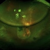
 Limited Choroidal Hemorrhage S/P Glaucoma Valve Implant OS; Retinoschisis
Aug 21 2023 by Angela Rico
A 52 year old Female presents to office S/P Glaucoma Valve Implant with IOP: 5mmHg OS
Photographer: Angela Rico M.D.
Condition/keywords: choroidal hemorrhage, glaucoma, hypotony, retinoschisis
-
 Suprachoroidal Hemorrhage
Sep 2 2020 by Rinal Pandit
Fundus photograph of left eye of a 56-year-old female with primary angle closure glaucoma showing massive hemorrhagic choroidal detachment that developed following trabeculectomy surgery. Suprachoroidal hemorrhage is defined as the accumulation of blood within the potential space between the choroid and sclera, with the source of the blood being the long or short posterior ciliary artery. Delayed suprachoroidal hemorrhage (DSHC) remains one of the most dreaded and sight threatening complications of glaucoma filtration surgery. The risk factors include old age, hypertension, high myopia, arteriosclerosis, chronically elevated IOP, sudden hypotony, trauma, aphakia/pseudophakia, prior vitrectomy, history of 5 FU injections and anti-platelet agents. The incidence of postoperative SCH after trabeculectomy varies between 0.6%- 1.4%. DSCH after surgery varies considerably in severity but is generally characterized by the sudden onset of severe pain, decreased vision, and a shallow anterior chamber usually associated with raised intraocular pressure. B-scan ultrasonography can help to distinguish serous from hemorrhagic choroidals.Suprachoroidal hemorrhages appear as dome-shaped elevations of the retina with increased echo densities that are often heterogeneous and within the suprachoroidal space. Choroidal effusions appear as dome-shaped elevations with hypoechoic suprachoroidal space. The first step in the management is the timely diagnosis. Medical management includes oral and topical antiglaucoma drugs to lower IOP, oral and topical steroids to control inflammation and topical cycloplegics and oral analgesics to tackle pain. Serial ultrasound B scans of the affected eye should be performed in order to monitor progression of the SCH and help determine apposition, height, and liquefaction of the SCH. Indications of surgical drainage include non resolution with medical management,concurrent retinal detachment, central retinal apposition (kissing choroidals) and incarceration of vitreous in the wound site. The ideal time of drainage is between 7-14 days depending upon clot lysis. The prognosis of both intraoperative and postoperative SCH is poor. An overwhelming majority of patients do not achieve pre-hemorrhage visual acuity and most do not recover to a visual acuity of 20/200 or better. The major determinants of good or bad visual outcomes of SCH’s are preoperative visual acuity and retinal detachment at the time of hemorrhage, respectively.
Imaging device: OPTOS,Ultra wide field retinal imaging system
Condition/keywords: suprachoroidal hemorrhage, trabeculectomy, ultra-wide field imaging
-
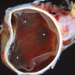 Enucleated Eye with Subretinal Hematoma
May 18 2020 by McGill University Health Centre
This enucleation specimen shows a diffuse and extended choroidal hemorrhage (*). Note the detached retina and extraocular hemorrhage (arrow).
Condition/keywords: subretinal hemorrhage
-
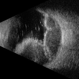 Suprachoroidal Hemorrhage
Nov 16 2019 by Sophia El Hamichi, MD
Ultrasound of the right eye of 43-year-old male presenting with suprachoroidal hemorrhage, note the multilobulated heterogenous echogenic mass aspect of the choroid
Photographer: Fiona J Ehlies, Murray Ocular Oncology and Retina, Miami
Condition/keywords: B scan ultrasound, suprachoroidal hemorrhage
-
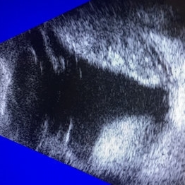 B-Scan Ultrasonography of a Suprachoroidal Hemorrhage
Oct 2 2019 by Radwan S. Ajlan, MBBCh, FRCS(C)
B-scan ultrasonography of a suprachoroidal hemorrhage.
Condition/keywords: B scan ultrasound, suprachoroidal hemorrhage
-
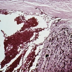 Slide 7-108
Feb 25 2019 by Lancaster Course in Ophthalmology
Expulsive hemorrhage. The blood lifts the choroid away from the sclera.
Condition/keywords: choroid, expulsive choroidal hemorrhage, sclera
-
 Choroidal Blow-Out
Jun 29 2018 by Gareth Lema, MD, PhD
Severe choroidal blow out caused by a suprachoroidal hemorrhage. There was a dense VH, and ultrasound showed a possible tear in the mid-periphery. Intraoperatively, a severe choroidal rupture secondary to a suprchoroidal hemorrhage was found temporally and a separate rhegmatogenous retinal detachment was found nasally. This photo was taken after silicone oil had been removed. The initial cause of injury was from a hockey ball in a young man.
Photographer: Sandra Boglione, Ross Eye Institute, University at Buffalo, Jacobs School of Medicine, Buffalo, NY
Imaging device: Optos
Condition/keywords: choroidal rupture
-
 Surgical Management of Massive Suprachoroidal Hemorrhage: Don’t Play It Blind!
Apr 6 2018 by Jay Sheth
Expulsive suprachoroidal hemorrhage(SCH) is a catastrophic complication of intraocular surgery. Current management includes SCH drainage through external sclerotomies & intermittent fundus evaluation by IO. We describe a novel surgical technique, utilizing chandelier-assisted wide-angled visualization of various steps of SCH drainage in 62/M. Using the wide-angle viewing system & 23G extrusion cannula through asclerotomy, active drainage of SCH was performed whereby we beautifully demonstrate separation of the kissing choroids with gradual unmasking of macula &disc underneath. Post-operatively, patient improved to CF3m & eye was successfully salvaged. Our educational video demonstrates that chandelier-assisted controlled drainage of SCH under continuous visualization is an easy technique to achieve excellent anatomical & visual outcomes with better safety profile. It can be instrumental in training residents & fellows who can simultaneously visualize surgical steps along with surgeon.
Condition/keywords: suprachoroidal hemorrhage, video
-
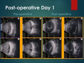
 POD 1
POD 1
Jan 7 2018 by John S. King, MD
61-year-old developed a large choroidal OD after a bleb revision. 30 years ago had an IOFB removed. The initial photos, POD 1 and POW 3 photos are provided. Dr. Zocchi performed the surgery. Surgery included 42 band placed 360 (superior bleb was fibrosed and left in place during conj peritomy); suprachoroidal drainiage was via radial incision; a pre-existing ST tear was present; the retinal adhesions were removed with blunt dissection, then remaining pvr was peeled; PFO used to flatten retina; 5000 cs sil oil used. Post-op vision has improved for near to J5.
Imaging device: Optos
Condition/keywords: suprachoroidal hemorrhage
-
 Pre-OP
Jan 7 2018 by John S. King, MD
61-year-old developed a large choroidal OD after a bleb revision. 30 years ago had an IOFB removed. The initial photos, POD 1 and POW 3 photos are provided. Dr. Zocchi performed the surgery. Surgery included 42 band placed 360 (superior bleb was fibrosed and left in place during conj peritomy); suprachoroidal drainiage was via radial incision; a pre-existing ST tear was present; the retinal adhesions were removed with blunt dissection, then remaining pvr was peeled; PFO used to flatten retina; 5000 cs sil oil used. Post-op vision has improved for near to J5.
Imaging device: Optos
Condition/keywords: suprachoroidal hemorrhage
-
 POW 3
POW 3
Jan 7 2018 by John S. King, MD
61-year-old developed a large choroidal OD after a bleb revision. 30 years ago had an IOFB removed. The initial photos, POD 1 and POW 3 photos are provided. Dr. Zocchi performed the surgery. Surgery included 42 band placed 360 (superior bleb was fibrosed and left in place during conj peritomy); suprachoroidal drainiage was via radial incision; a pre-existing ST tear was present; the retinal adhesions were removed with blunt dissection, then remaining pvr was peeled; PFO used to flatten retina; 5000 cs sil oil used. Post-op vision has improved for near to J5.
Imaging device: Optos
Condition/keywords: suprachoroidal hemorrhage
-
 Choroidal Hemorrhage, Subretinal Hemorrhage
Dec 18 2017 by Nichole Lewis
Choroidal hemorrhage, Subretinal Hemorrhage, wet macular degeneration,
Photographer: Nichole Lewis
Condition/keywords: choroidal hemorrhage, choroidal neovascularization (CNV), exudative age-related macular degeneration, subretinal hemorrhage, wet age-related macular degeneration (wet AMD)
-
 Superchoroid Hemorrhage
Feb 12 2015 by H. Michael Lambert, MD
A-scan shows choroidal detachment with hemorrhage.
Condition/keywords: choroidal detachment, choroidal hemorrhage
-
 Superchoroid Hemorrhage
Feb 12 2015 by H. Michael Lambert, MD
Choroidal detachment.
Condition/keywords: choroidal detachment, choroidal hemorrhage
-
 Superchoroid Hemorrhage
Feb 12 2015 by H. Michael Lambert, MD
Choroid detachment with vitreous hemorrhage.
Condition/keywords: choroidal detachment, choroidal hemorrhage
-
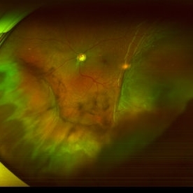
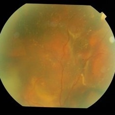 Massive Suprachoroidal & Subretinal Hemorrhage in IPCV
Oct 7 2014 by Mallika Goyal, MD
Fundus photograph of the right eye of a 53-year-old lady with massive subretinal and suprachoroidal hemorrhage 4 weeks after PDT and 1 week after Avastin for IPCV.
Photographer: Mallika Goyal, MD, Apollo Health City, Jubilee Hills, Hyderabad, India
Condition/keywords: suprachoroidal hemorrhage
-
 Massive Suprachoroidal & Subretinal Hemorrhage in IPCV
Oct 7 2014 by Mallika Goyal, MD
Fundus photograph of the right eye of a 53-year-old lady with massive subretinal and suprachoroidal hemorrhage 4 weeks after PDT and 1 week after Avastin for IPCV.
Photographer: Mallika Goyal, MD, Apollo Health City, Jubilee Hills, Hyderabad, India
Condition/keywords: suprachoroidal hemorrhage
 Loading…