Search results (261 results)
-
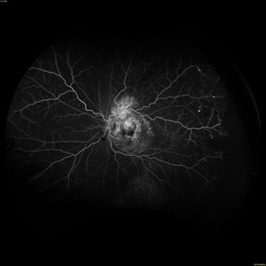
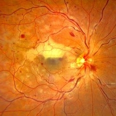
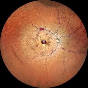 Ultra-Wide Field Image of Central Retinal Vein Occlusion with Foveal Hemorrhage
Apr 17 2025 by Malvika Singh
Ultra- wide field fundus photograph of a 41 year-old male, with a central retinal vein occlusion and a foveal sub-internal limiting membrane hemorrhage.
Photographer: Dr Malvika Singh, Retina Foundation, Ahmedabad, India
Imaging device: Mirante SLO/OCT
Condition/keywords: central retinal vein occlusion (CRVO), macular hemorrhage, Ultra-wide field retinal imaging
-
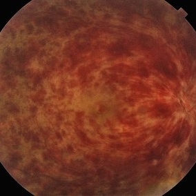
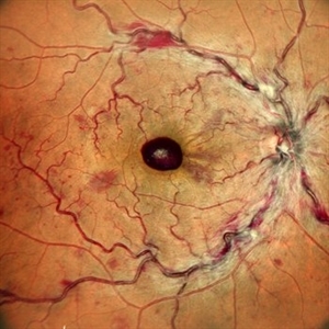 Central Retinal Vein Occlusion with Foveal Hemorrhage
Apr 17 2025 by Malvika Singh
Fundus photograph of a 41 year-old, male, with a central retinal vein occlusion and a foveal sub-internal limiting membrane hemorrhage.
Photographer: Dr Malvika Singh, Retina Foundation, Ahmedabad, India
Imaging device: Mirante SLO/OCT
Condition/keywords: central retinal vein occlusion (CRVO), macular hemorrhage
-
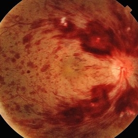
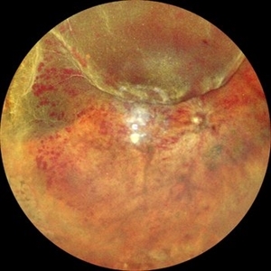 CRVO with RD
Apr 4 2025 by Tejaswita Verma
Fundus photo of a 58 year-old hypertensive male who presented with RE 6/60 vision with CRVO, rhegmatogenous RD.H/O DOV in RE since 3 days, H/O receiving anti VEGF X 2 injections 2 months ago
Photographer: DR. TEJASWITA VERMA
Imaging device: MIRANTE
Condition/keywords: central retinal vein occlusion (CRVO), Retinal Detachment
-
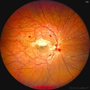
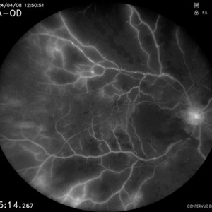
 Shunt Vessels
Apr 1 2025 by Korey Starkey
62-year-old patient presented with stable CRVO in the left eye. FA performed that day shows delayed AV transit is present, this has compensated with shunt vessels at the disc. However there is no evidence of active leakage. OS vision 20/25.
Photographer: Korey Starkey
Imaging device: Topcon
Condition/keywords: central retinal vein occlusion (CRVO), fundus photograph, optic nerve, shunts vessels, Topcon
-
 Central Retinal Vein Occlusion with Macular Edema
Jan 29 2025 by Kimberly Wakester
Fundus photograph of a 62-year-old man with central retinal vein occlusion with macular edema and a new PVD with an operculated retinal tear in the left eye. Laser to retinal tear was completed. Patient will return in 2-3 weeks for follow up exam with possible intravitreal injection for the CRVO with edema and to follow up on the operculated retinal tear s/p retinal tear laser.
Photographer: Kimberly Wakester, COA
Imaging device: Optos California
Condition/keywords: central retinal vein occlusion (CRVO), operculated tear, PVD
-
 CRVO
CRVO
Jan 15 2025 by Virginia Gebhart
65 year old male with new central retinal vein occlusion with macular edema. Carotid ultrasound showed less than 50% stenosis bilateral. Dilated and tortuous vessels as well as cystoid macular edema and flame-shaped hemes in all 4 quadrants. Treated with IVA
Photographer: Virginia Gebhart, Retina Consultants of Carolina
Imaging device: Optos California
Condition/keywords: central retinal vein occlusion (CRVO), macular edema
-
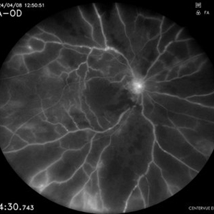
 Chronic CRVO
Dec 12 2024 by Korey Starkey
Fluorescein Angiography of a 62 year-old man with chronic central retinal vein occlusion. Vision is 20/200.
Photographer: Korey Starkey
Imaging device: Optos
Condition/keywords: capillary nonperfusion, central retinal vein occlusion (CRVO), FLUORESCEIN ANGIOGRAPHY, ischemia, microaneurysms, Optos
-
 Central Retinal Vein Occlusion
Sep 27 2024 by Korey Starkey
Fluorescein angiogram of a 75 year old patient with central retinal vein occlusion. FA shows areas of patchy ischemia and petaloid leakage. Patient is being treated with anti-vegf treatments at this time.
Photographer: Korey Starkey
Condition/keywords: central retinal vein occlusion (CRVO), FLUORESCEIN ANGIOGRAPHY, ischemia, macular edema, petaloid leakage, ultra-widefield image
-
 Central Retinal Vein Occlusion
Sep 27 2024 by Jeffrey Barker
65 year old male with a Central Retinal Vein Occlusion and Macular Edema and Capillary Nonperfusion.
Photographer: Jeffrey P. Barker
Condition/keywords: central retinal vein occlusion (CRVO), macular edema
-
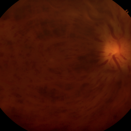
 Ischemic Central Retinal Vein Occlusion
Aug 6 2024 by César Adrián Gómez Valdivia, MD
Fundus photograph of an 80 year old man with and acute central retinal vein occlusion, ischemic variant.
Photographer: @eyemissu2
Condition/keywords: central retinal vein occlusion (CRVO), ischemic CRVO
-
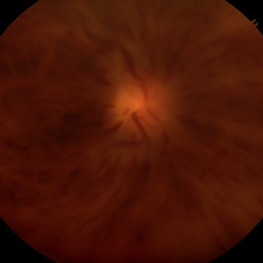
 Central Retinal Vein Occlusion
Jul 21 2024 by César Adrián Gómez Valdivia, MD
Central Retinal Vein Occlusion found in a 72 year old patient with history of uncontrolled Hypertension. Non-Ischemic Variant.
Photographer: Erika Paulina Ornelas Cazares
Imaging device: TOPCON TRC-50DX
Condition/keywords: central retinal vein occlusion (CRVO)
-
 Central Retinal Vein Occlusion with Macular Edema OS
Jul 5 2024 by Zach Seim
Optos fundus photograph of a Central Retinal Vein Occlusion in a 20 year old male. Vision at presentation was Dsc 20/25-1.
Photographer: Zach Seim
Imaging device: Optos California
Condition/keywords: central retinal vein occlusion (CRVO), left eye, macular edema, Optos, OPTOS CALIFORNIA
-
 Combined Central Retinal Vein Occlusion with Branch Retinal Artery Occlusion
Apr 29 2024 by KANWALJEET HARJOT MADAN, M.S. (Ophthalmology), FAICO (Vitreous - Retina)
This is fundus photograph of a 46-year male patient who presented with sudden diminution of vision in his right eye (RE) for 3 days. He was hypertensive but non diabetic. On examination, his best corrected vision in RE was 6/12. His left eye (LE) was normal. His fundus examination in RE revealed multiple intra retinal hemorrhages in all quadrants with tortuosity of veins suggestive of central retinal vein occlusion (CRVO) with mild disc edema. An ischemic area was seen superior to fovea suggestive of branch retinal artery occlusion. OCT depicted thickening of inner retinal layers with little evidence of macular edema. Hematological and cardio vascular investigations were done. He had bilateral thickening of intimal and medial walls of carotid arteries. He was under cardiology treatment. His vision improved to 6/6.
Photographer: Dr. Kanwaljeet Harjot Madan, M.S. (Ophthalmologist) Fellow in Vitrous & Retina. Thind Eye Hospital, Jalandhar City. Punjab. India
Condition/keywords: branch retinal artery occlusion (BRAO), central retinal vein occlusion (CRVO)
-
 Combined Central Retinal Vein Occlusion with Branch Retinal Artery Occlusion
Apr 28 2024 by KANWALJEET HARJOT MADAN, M.S. (Ophthalmology) Fellow in Vitreous & Retina
This is fundus photograph of a 46-year male patient who presented with sudden diminution of vision in his right eye (RE) for 3 days. He was hypertensive but non diabetic. On examination, his best corrected vision in RE was 6/12. His left eye (LE) was normal. His fundus examination in RE revealed multiple intra retinal hemorrhages in all quadrants with tortuosity of veins suggestive of central retinal vein occlusion (CRVO) with mild disc edema. An ischemic area was seen superior to fovea suggestive of branch retinal artery occlusion. OCT depicted thickening of inner retinal layers with little evidence of macular edema. Hematological and cardio vascular investigations were done. He had bilateral thickening of intimal and medial walls of carotid arteries. He was under cardiology treatment. His vision improved to 6/6.
Photographer: Dr Kanwaljeet Harjot Madan
Condition/keywords: branch retinal artery occlusion (BRAO), central retinal vein occlusion
-
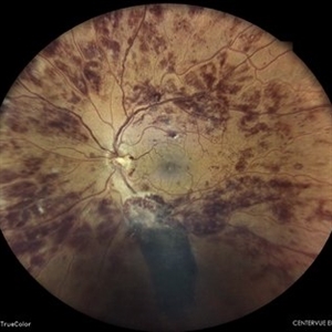 Central Retinal Vein Occlusion
Apr 9 2024 by Akansha Sharma
Color fundus photograph of a 73 year old hypertensive male with central retinal vein occlusion.
Photographer: Dr. Akansha Sharma, Bharati Eye Hospital
Condition/keywords: central retinal vein occlusion (CRVO), ischemic CRVO
-
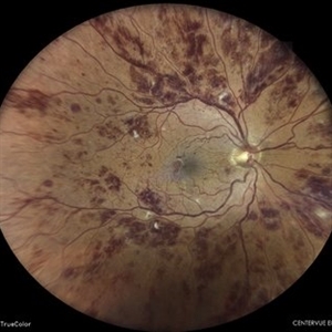 Central Retinal Vein Occlusion
Apr 9 2024 by Akansha Sharma
Color fundus photograph of a 73 year old hypertensive male with central retinal vein occlusion.
Photographer: Dr. Akansha Sharma, Bharati Eye Hospital
Condition/keywords: central retinal vein occlusion (CRVO), ischemic CRVO
-
 Combined Central Retinal Artery and Vein Occlusion
Apr 8 2024 by Akansha Sharma
Color fundus photograph of a 63 year old male with combined central retinal artery and vein occlusion with carotid artery stenosis and infarct in the brain.
Photographer: Dr. Akansha Sharma, Bharati Eye Hospital
Condition/keywords: central retinal artery occlusion (CRAO), central retinal vein occlusion (CRVO), CRAO
-
 Combined Central Retinal Artery and Vein Occlusion
Apr 8 2024 by Akansha Sharma
Fundus fluorescein angiography of a 63 year old male with combined central retinal artery and vein occlusion with carotid artery stenosis and infarct in the brain demonstrating late filling.
Photographer: Dr. Akansha Sharma, Bharati Eye Hospital
Condition/keywords: central retinal artery occlusion (CRAO), central retinal vein occlusion (CRVO), CRAO
-
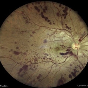
 Central Retinal Vein Occlusion
Jan 28 2024 by Gayathri Mohan
Fundus photograph of a 50 year old woman showing a CRVO.
Photographer: Dr Gayathri Mohan, Thumbay Medical and Dental Speciality Centre, U.A.E
Imaging device: 3nethra
Condition/keywords: central retinal vein occlusion (CRVO), ischemic CRVO
-
 Central Retinal Vein Occlusion with Macular Edema in Antiphospholipid Syndrome
Dec 24 2023 by Nikhil K Bommakanti, MD
A man in his thirties presented with a central retinal vein occlusion with macular edema in the right eye. Vision improved from 20/70 to 20/25 after 1 treatment with intravitreal bevacizumab. Laboratory testing revealed the presence of lupus anticoagulant.
Condition/keywords: antiphospholipid antibody syndrome, central retinal vein occlusion (CRVO), cystoid macular degeneration, macular edema
-
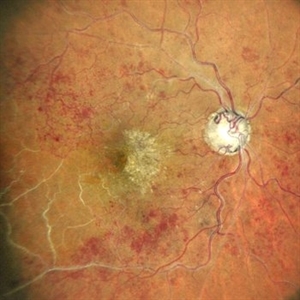 Central vein occlusion (chronic)
Nov 26 2023 by Anjana Mirajkar, MS Ophthalmology
A central color photo image of RE of a 60 year old male in a case of central vein occlusion.
Photographer: Dr. Anjana Mirajkar -Retina Foundation, Ahmedabad
Imaging device: Mirante-Nidek
Condition/keywords: central retinal vein occlusion (CRVO), ischemic CRVO
-
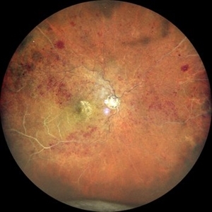 Central vein occlusion (chronic)
Nov 26 2023 by Anjana Mirajkar, MS Ophthalmology
A widefield color photo image of RE of a 60 year old male in a case of central vein occlusion.
Photographer: Dr. Anjana Mirajkar -Retina Foundation, Ahmedabad
Imaging device: Mirante-Nidek
Condition/keywords: central retinal vein occlusion (CRVO), central vein occlusion, ischemic CRVO
 Loading…