Search results (222 results)
-
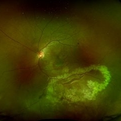
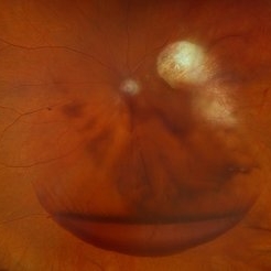
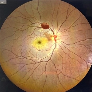
 Choroidal Rupture
Dec 4 2025 by Vishal Agrawal, MD, FRCS,FACS,FASRS
Color fundus pic of Left Eye showing curvilinear, crescent-shaped choroidal rupture, extending across the macular region, post blunt trauma. The rupture appears bright, yellow-white streak with well-delineated margins, consistent with exposed underlying sclera due to disruption of the choriocapillaris and Bruch's membrane.
Photographer: Dr Ayushi Gupta, Agrawal Hospital, Jaipur
Imaging device: Clarus 700
Condition/keywords: blunt trauma, choroidal rupture
-
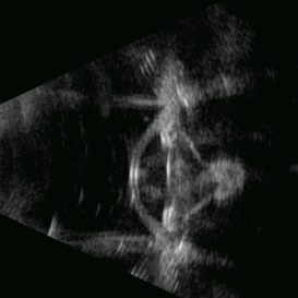
 Closed Funnel RD
Sep 26 2025 by Virginia Gebhart
B scan ultrasound of 12 year old male with complete closed funnel RD. Pt endorses several prior blunt traumas which may have caused detachment. If not secondary to trauma, may represent end-stage Coat's disease. FEVR unlikely due to FA findings of fellow eye. Eye is stable since initial visit in 2024, surgical intervention not recommended at this time. Vision NLP
Photographer: Virginia Gebhart, Retina Consultants of Carolina
Imaging device: Keeler Accutome
Condition/keywords: B scan ultrasound, chronic retinal detachment, Closed funnel RD, retinal detachment
-
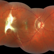
 Post Blunt Trauma Posterior Break
Aug 13 2025 by Debarun Sharma
Fundus photograph of a 21 year old male with history of blunt trauma with fist presenting with a large linear posterior break just adjacent to the infero-temporal arcade with choroidal rupture and subretinal bleed passing through fovea. Successful barrage laser of the break can be seen.
Photographer: Debarun Sharma, Sri Sankardeva Nethralaya, Guwahati
Imaging device: Optos
Condition/keywords: blunt trauma, full thickness retinal tear, laser photocoagulation
-
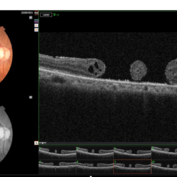 Double Macular Holes
Jun 26 2025 by Moazzam Parvez
OCT image of a 62 year old man after a blunt trauma by a tennis ball with a vision of CF 3 mt in the right eye.
Photographer: Moazzam Parvez , Netralayam , Kolkata
Imaging device: Topcon Maestro 2
Condition/keywords: double, traumatic macular hole
-
 Post-traumatic Choroidal Rupture
Jun 20 2025 by Alexander Babaev
Fundus photograph of a 46-year-man with a choroidal rupture after blunt trauma, complicated CNV.
Photographer: Babaev Alexander, Saint-Petersburg, medical clinic "Vision"
Imaging device: Carl Zeiss Visucam 500
Condition/keywords: choroidal rupture
-
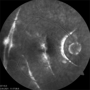 Post-traumatic Choroida Rupture-Fluorescein Angiography
Jun 20 2025 by Alexander Babaev
Fluorescein angiography of an 46-year-man with a choroidal rupture after blunt trauma, complicated CNV. 07.15, Dye leakage is visible along the edges of the rupture
Photographer: Babaev Alexander, Saint-Petersburg, medical clinic "Vision"
Imaging device: Carl Zeiss Visucam 500
Condition/keywords: blunt trauma
-
 Post-traumatic Choroidal Rupture-Fluorescein Angiography
Jun 20 2025 by Alexander Babaev
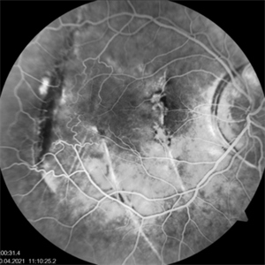
Fluorescein angiography of an 46-year-man with a choroidal rupture after blunt trauma, complicated CNV. 00.31s, Dye leakage is visible along the edges of the rupture
Photographer: Babaev Alexander, Saint-Petersburg, medical clinic "Vision"
Imaging device: Carl Zeiss Visucam 500
Condition/keywords: fluorescein angiogram (FA)
-
 Post-traumatic Choroidal Rupture-Fluorescein Angiography
Jun 20 2025 by Alexander Babaev
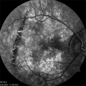
Fluorescein angiography of an 46-year-man with a choroidal rupture after blunt trauma, complicated CNV. 00.16s
Photographer: Babaev Alexander, Saint-Petersburg, medical clinic "Vision"
Imaging device: Carl Zeiss Visucam 500
Condition/keywords: blunt trauma
-
 Traumatic T
May 5 2025 by Gustavo Uriel Fonseca Aguirre
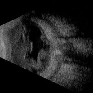
This B-mode axial ultrasound scan reveals vitreous hemorrhage, a folded retinal detachment, and sub-Tenon’s fluid extending into the optic nerve sheath, forming the characteristic 'T-sign.' These findings are consistent with severe posterior segment trauma secondary to blunt ocular injury.
Photographer: Gustavo U. Fonseca Aguirre, Hospital Conde de Valenciana, Ciudad de México
Condition/keywords: blunt trauma, retinal detachment, T sign
-
 Traumatic Retinal Detachment
May 5 2025 by Gustavo Uriel Fonseca Aguirre
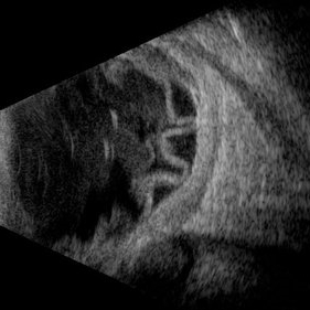
This B-mode longitudinal ultrasound scan over the macular area reveals vitreous hemorrhage, retinal detachment with folding, peripheral annular choroidal detachment, and sub-Tenon's fluid in the setting of blunt ocular trauma. The findings indicate severe posterior segment disruption with multi-compartment involvement.
Photographer: Gustavo U. Fonseca Aguirre, Hospital Conde de Valenciana, Ciudad de México
Condition/keywords: blunt trauma, Retinal Detachment
-
 Hourglass in an Eye
Apr 22 2025 by KRISHNENDU NANDI, MS
A twenty-five-year-young male presented with a decrease in vision in the right eye following a blunt trauma with a football. On examination the BCVA in the right eye was CFCF and the left eye was 6/6, N6. The anterior segment was within normal limits. AT was 12 and 10 mm of Hg in the right and left eyes, respectively. Fundus examination reveals subhyaloid haemorrhage in the right eye with an attached retina. The fundus of the left eye was within normal limits. YAG laser hyaloidotomy was done with an energy of 2 mJ in the right eye. After 3 weeks the BCVA in the right eye improved to 6/9, N6.
Photographer: Dr. Krishnendu Nandi
Imaging device: Topcon
Condition/keywords: Trauma, YAG HYALOIDOTOMY, Young Male
-
 Traumatic Hemorrhage
Apr 17 2025 by Virginia Gebhart
60 year old male with vitreous and sub hyaloid hemorrhage from being hit in the eye. No holes, tears, or detachment. Will observe closely, if no improvement will consider surgical repair. Treated melanoma s/p brachytherapy in 2008.
Photographer: Virginia Gebhart, Retina Consultants of Carolina
Imaging device: Optos California
Condition/keywords: blunt trauma, sub hyaloid hemorrhage, vitreous hemorrhage
-
 Traumatic Posterior Capsular Rupture
Apr 9 2025 by Gustavo Uriel Fonseca Aguirre
Immersion B-mode ultrasound in a patient with blunt ocular trauma demonstrates an isolated posterior lens capsule rupture accompanied by phacodonesis.
Photographer: Gustavo U. Fonseca Aguirre, Hospital Conde de Valenciana, Ciudad de México
Condition/keywords: blunt trauma, Posterior Capsular Rupture
-
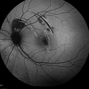 Choroidal Rupture
Apr 7 2025 by Ramses Rosales-Diaz
Autofluorescence image of a 39-year-old female patient who sustained blunt ocular trauma resulting in three choroidal ruptures.
Photographer: Ramses Rosales-Diaz, Asociación Para Evitar la Ceguera en México I.A.P., Mexico City
Imaging device: Heidelberg Spectralis
Condition/keywords: blunt trauma, Choroidal Rupture
-
 Intraocular Foreign Body
Apr 3 2025 by Gustavo Uriel Fonseca Aguirre
B-mode ultrasonography of an eye with a 1-year history of suspected blunt trauma revealed an incidental intraocular foreign body within the vitreous cavity.
Photographer: Gustavo U. Fonseca Aguirre, Hospital Conde de Valenciana, Ciudad de México
Condition/keywords: intraocular foreign body
-
 Firework Injury
Feb 13 2025 by Virginia Gebhart
44 year old male presented New Year's Day for trauma after fireworks injury. Choroidal rupture temporal macula, inferior vitreous hemorrhage, and extensive RPE changes in the macula. Significant improvement since initial presentation. Limited central vision, guarded prognosis due to extensive blunt trauma.
Photographer: Virginia Gebhart, Retina Consultants of Carolina
Imaging device: Optos California
Condition/keywords: blunt trauma, choroidal rupture, commotio retinae, firework injury, secondary glaucoma, subretinal hemorrhage, VH, vitreous hemorrhage
-
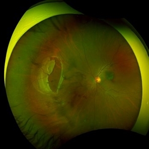
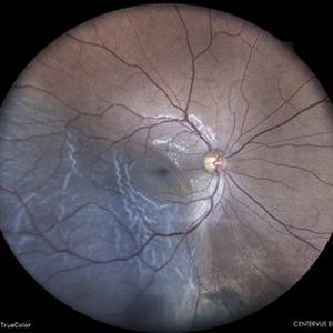
 A Large Break at the Posterior Pole With RD With PVR (S/p Old Blunt Trauma)
Jan 16 2025 by Anand Temkar
Right eye widefield fundus color photo of a 10 year old kid who noticed diminution of vision in right eye since a month. We can see the large break at the posterior pole with rolled up margins associated with retinal detachment and PVR changes.
Photographer: Dr.Anand Temkar- Retina Foundation, Ahmedabad
Imaging device: Mirante
Condition/keywords: posterior pole break, proliferative vitreoretinopathy (PVR), Retinal Detachment
-
 A Large Break at the Posterior Pole With RD With PVR (S/p Old Blunt Trauma)
Jan 16 2025 by Anand Temkar
Right eye central fundus color photo of a 10 year old kid who noticed diminution of vision in right eye since a month. We can see the large break at the posterior pole with rolled up margins associated with retinal detachment and PVR changes.
Photographer: Dr.Anand Temkar- Retina Foundation, Ahmedabad
Imaging device: Mirante
Condition/keywords: Posterior pole break, proliferative vitreoretinopathy (PVR), Retinal Detachment
-
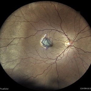
 Traumatic Macular Hole pre and post repair
Nov 25 2024 by Shobhit Chawla, M.S.
31 year-old male reported with h/o of blunt trauma over right eye ,from cricket ball. On examination DVA RE 6/60,LE 6/18,ant segment BE :WNL,FUNDUS RE:Sub retinal hemorrhage at macula with chroidal tear,LE :WNL. Undwer went 25G vitrectomy+sub retinal TPA+C3F8(RE).Post op 1 month DVA RE:6/24 ,ANT SEGMENT:WNL,FUNDUS:resolved sub retinal haem with traumatic macular hole. Under went repeat vit+autologous retinal transplant +SOI RE.POST SOR AFTER4monthsV/A :6/18 RE
Photographer: Ranjit Ray
Imaging device: Clarus 500
Condition/keywords: Macular hole, retinal graft, subretinal hemorrhage
-
 Large Retinal Tear from a Shuttlecock Injury
Oct 11 2024 by Ahmad B. Tarabishy, MD
27 year old woman presenting with floaters and a shadow in her temporal visual field OS. Approximately one week earlier, she was injured in her left eye by a shuttlecock while playing badminton. Fundus exam reveals mild vitreous hemorrhage and a large retinal tear with a small cuff of surrounding SRF. This image was taken immediately following treatment with barrier laser retinopexy.
Photographer: Angela Rico, M.D.
Imaging device: Optos
Condition/keywords: blunt trauma, ocular trauma, retinal tear
-
 Large Retinal Tear from a Shuttlecock Injury
Oct 11 2024 by Ahmad B. Tarabishy, MD
27 year old woman presenting with floaters and a shadow in her temporal visual field OS. Approximately one week earlier, she was injured in her left eye by a shuttlecock while playing badminton. Fundus exam reveals mild vitreous hemorrhage and a large retinal tear with a small cuff of surrounding SRF.
Photographer: Angela Rico, M.D.
Imaging device: Optos
Condition/keywords: blunt trauma, ocular trauma, retinal tear
-
 Traumatic CRAO with Cilioretinal Artery Sparing
Sep 10 2024 by KRISHNENDU NANDI, MS
A 25 year-old male presented with dimness of vision in the right eye for the last 3 days following blunt trauma. The BCVA of the right eye was CF close to the face and left eye was 6/6, N6 in Snellen’s chart. On examination the retina showed CRAO with cherry red spot and sparing of cilioretinal artery circulation. Traumatic subhyaloid haemorrhage also noted at supero-temporal arcade.
Photographer: Dr Krishnendu Nandi
Condition/keywords: CRAO, subhyaloid hemorrhage, Trauma
-
 Berlin’s Edema
Aug 10 2024 by Sachit Mahajan, MBBS MS
Fundus photograph of 10 year old boy, with a history of blunt trauma to left eye with cricket ball in school, showing Berlin’s Edema at posterior pole.
Photographer: Prattoy, Dr Shroff’s Charity Eye Hospital, New Delhi
Imaging device: Mirante, Nidek
Condition/keywords: Berlin's edema, blunt trauma, ocular trauma
-
 Subretinal Hemorrhage Status Post Blunt Trauma
May 27 2024 by Akansha Sharma
Color fundus photograph of a 41 year old male with subretinal bleed status post blunt trauma.
Photographer: Dr. Akansha Sharma, Bharati Eye Hospital
Condition/keywords: subretinal hemorrhage, trauma
-
 Retinal Detachment
May 27 2024 by Akansha Sharma
Color fundus photograph of a 30 year old male with retinal detachment status post blunt trauma.
Photographer: Dr. Akansha Sharma, Bharati Eye Hospital
Condition/keywords: RD, Retinal Detachment
 Loading…