Search results (25 results)
-
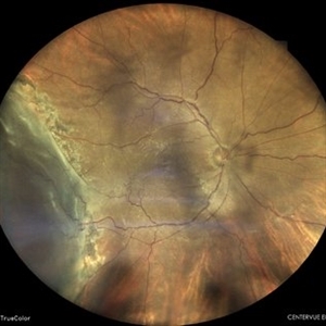
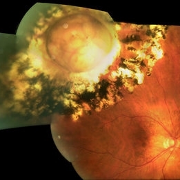
 Large Retinal Tear from a Shuttlecock Injury
Oct 11 2024 by Ahmad B. Tarabishy, MD
27 year old woman presenting with floaters and a shadow in her temporal visual field OS. Approximately one week earlier, she was injured in her left eye by a shuttlecock while playing badminton. Fundus exam reveals mild vitreous hemorrhage and a large retinal tear with a small cuff of surrounding SRF. This image was taken immediately following treatment with barrier laser retinopexy.
Photographer: Angela Rico, M.D.
Imaging device: Optos
Condition/keywords: blunt trauma, ocular trauma, retinal tear
-
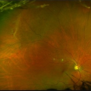
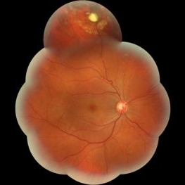
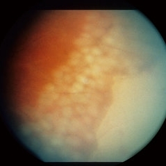
 Choroidal Osteoma
Jun 13 2024 by Virginia Gebhart
20 year old female with choroidal osteoma. Stable s/p PDT x3 and focal laser x 2, no obvious progression on last exam. Monitoring closely. Vision 20/30.
Photographer: Virginia Gebhart
Imaging device: Topcon 50 DX
Condition/keywords: barrier laser, choroidal osteoma, PDT
-
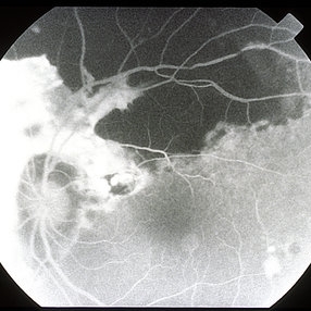
 FAF of Barricade Laser on Choroidal Osteoma
Jun 12 2024 by Virginia Gebhart
20 year old female with stable choroidal osteoma s/p PDT x 3 and focal laser x 2. No obvious progression on last exam, vision 20/30. Monitoring closely.
Photographer: Virginia Gebhart
Imaging device: Topcon 50 DX
Condition/keywords: autofluorescence imaging, barrier laser, choroidal osteoma, focal laser, fundus autofluorescence (FAF)
-
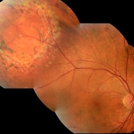
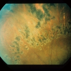
 RETINAL DETACHMENT STATUS POST BARRAGE LASER FOR HORSE SHOE TEAR
May 31 2023 by Akansha Sharma
COLOUR FUNDUS PHOTOGRAPH OF A 58 YEAR OLD MALE PATIENT WITH RETINAL DETACHMENT STATUS POST BARRAGE LASER FOR HORSE SHOE TEAR
Photographer: Dr. Urmil Shah, Dr. Akansha Sharma, Dr. Denish Patel,
Condition/keywords: barrier laser, Retinal Detachment
-
 Bullous Retinoschisis with Outer Retinal Holes
Jun 15 2020 by Olivia Rainey
Ultra-widefield pseudocolor fundus photograph of a 56-year-old female with bullous retinoschisis with outer retinal holes affecting her right eye. The physician noted superotemporal retinoschisis in her monoculcar functioning eye. There was no demarcation line and no inner or outer layer breaks at her first appointment in February of 2020. On 6/15/20 she had a new onset outer holes and SRF tracking inferiorly. The physician recommended observation, however if this continues to progress we have discussed indications for barrier laser.
Photographer: Olivia Rainey, OCT-C, COA
Imaging device: Optos California
Condition/keywords: bullous retinoschisis, Optos, outer layer breaks, outer layer hole, pseudocolor, subretinal fluid, superior retina, ultra-wide field imaging
-
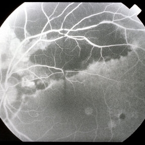
 Retinal Break
Feb 12 2020 by DIEGO TOLENTINO
Retinal break at vascular junction and laser barricade.
Photographer: Diego Tolentino, CEOP
Condition/keywords: barrier laser, retinal break
-
 Operculated Hole with Barrier Laser
Nov 5 2019 by Nichole Lewis
66-year-old female with an operculated retinal hole s/p barrier laser treatment. Choroidal Nevus Inferior.
Photographer: Nichole Lewis
Imaging device: Optos
Condition/keywords: barrier laser, choroidal nevus, operculated retinal hole, operculated tear
-
 Giant Retinal Tear
May 1 2018 by Talia R Kaden, MD
Fundus Photograph of the right eye of a 51-year-old taxi driver with a newly lasered giant retinal tear. He had a history of a retinal detachment from a giant retinal tear in his left eye.
Photographer: Maria Pei, Bellevue Hospital, New York University, NY
Imaging device: Topcon TRC 501x
Condition/keywords: barrier laser, fresh laser burns, giant retinal tear
-
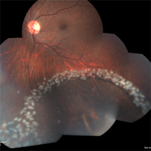
 Laser Barrage for Temporal Localized Rhematogenous Retinal Detachment
Feb 15 2018 by Kushal S Delhiwala, MBBS, MS, FMRF,FICO, FAICO
39-year-old female presenting with sudden onset flashes and floaters in left eye having undergone refractive surgery 20 years before for pathologic myopia.Color fundus photograph montage of left eye showing macula sparing inferotemporal localized Rhematogenous retinal detachment with horse shoe tear and temporal lattice degeneration treated with laser barrage.
Photographer: Dr Kushal Delhiwala, Netralaya superspeciality eye hospital ,Ahmedabad
Imaging device: Zeiss Visucam 500
Condition/keywords: barrier laser, macula sparring
-
 Scleral Ectasia With Barrier Laser
Sep 1 2017 by Nichole Lewis
Scleral ectasia with barrier laser.
Photographer: Nichole Lewis
Condition/keywords: scleral ectasia
-
 Macula-Sparing GRT RRD
Jul 6 2017 by Andrew A. Moshfeghi, MD, MBA, FASRS
Wide-field fundus photograph of a 43-year-old myopic man with a history of lattice retinal degeneration status posterior barrier laser performed elsewhere who presented with a giant-retinal tear associated retinal detachment of the right eye.
Photographer: Jay Jiang, University of Southern California Roski Eye Institute
Imaging device: Optos California
Condition/keywords: acute retinal detachment, giant retinal tear, lattice degeneration
-
 Retinal Detachment
May 13 2016 by Nichole Lewis
Inferior Retinal Detachment with some demarcation line s/p barrier laser.
Photographer: Nichole Lewis
Condition/keywords: barrier laser
-
 Retinal Detachment
May 9 2016 by Nichole Lewis
Retinal detachment with partial demarcation line and same day barrier laser treatment.
Photographer: Nichole Lewis
-
 Treated Retinal Tear
Apr 1 2016 by Nichole Lewis
Retinal tear with barrier laser.
Photographer: Nichole Lewis - Pennsylvania Retina Specialists, Camp Hill, PA
Condition/keywords: retinal tear
-
 Intraretinal Foreign Body
Oct 10 2015 by Hamid Ahmadieh, MD
Merged color fundus photograph of the right eye of a patient with intraretinal metallic foreign body . Barrier laser photocoagulation was performed before vitrectomy and foreign body removal.
Photographer: Soodabeh Fouladin, Negah Eye Center, Tehran, Iran
Condition/keywords: color fundus photograph, intraocular foreign body
-
 Lasered Retinal Break
May 2 2014 by Neha Goel, MS DNB FRCS (Glasg)
Montage image of the right eye of a 40-year-old male showing a large retinal tear temporal to the macula, that was lasered.
Photographer: Neha Goel
Imaging device: Zeiss Visucam
Condition/keywords: barrier laser, full thickness retinal tear
-
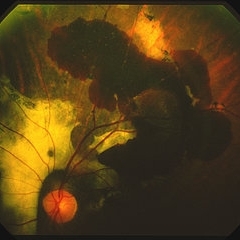
 Resolved ARN Post Barrier Laser
Jul 29 2013 by H. Michael Lambert, MD
Resolved ARN post barrier laser
Condition/keywords: acute retinal necrosis
-
 Resolved ARN Post Barrier Laser
Jul 29 2013 by H. Michael Lambert, MD
Resolved ARN post barrier laser
Condition/keywords: acute retinal necrosis
-
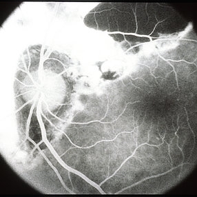
 ARN post barrier laser
Jul 29 2013 by H. Michael Lambert, MD
Acute retinal necrosis post barrier laser
Condition/keywords: acute retinal necrosis
-
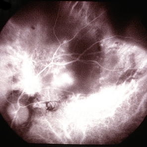
 Thick Subretinal Hemorrhage 10
Mar 14 2013 by Maurice F. Rabb
59-year-old black woman with a thick subretinal hemorrhage with vision 20/20. The barrier laser was extended, but the hemorrhage not treated.
Condition/keywords: 20/20, subretinal hemorrhage
-
 Thick Subretinal Hemorrhage 9
Mar 14 2013 by Maurice F. Rabb
59-year-old black woman with a thick subretinal hemorrhage with vision 20/20. The barrier laser was extended, but the hemorrhage not treated.
Condition/keywords: 20/20, subretinal hemorrhage
-
 Thick Subretinal Hemorrhage 7
Mar 14 2013 by Maurice F. Rabb
59-year-old black woman with a thick subretinal hemorrhage with vision 20/20. The barrier laser was extended, but the hemorrhage not treated.
Condition/keywords: 20/20, subretinal hemorrhage
-
 Thick Subretinal Hemorrhage 6
Mar 14 2013 by Maurice F. Rabb
59-year-old black woman with a thick subretinal hemorrhage with vision 20/20. The barrier laser was extended, but the hemorrhage not treated.
Condition/keywords: 20/20, subretinal hemorrhage
-
 Thick Subretinal Hemorrhage 11
Mar 14 2013 by Maurice F. Rabb
59-year-old black woman with a thick subretinal hemorrhage with vision 20/20. The barrier laser was extended, but the hemorrhage not treated.
Condition/keywords: 20/20, subretinal hemorrhage
-
 Post-Op Day 10 Barrier Laser
Feb 13 2013 by From the Collections of Thomas M. Aaberg, MD and Thomas M. Aaberg Jr., MD
Barrier laser, preretinal hemorrhage.
Condition/keywords: barrier laser, post-op, preretinal hemorrhage
 Loading…