Search results (27 results)
-
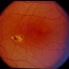
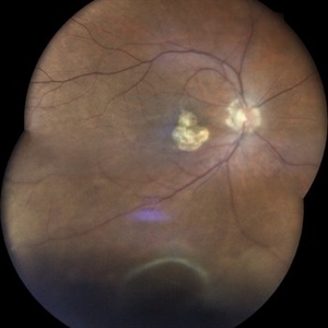 Scarred Choroidal Neovacular Membrane With Large Inferior Horse Shoe Tear
Feb 7 2024 by Akansha Sharma
Color fundus photograph of a 73 year old male with scarred choroidal neovascular membrane with large horse shoe tear inferiorly.
Photographer: Dr. Akansha Sharma, Bharati Eye Hospital
Condition/keywords: atrophic scar, choroidal neovascular membrane (CNVM), CNVM
-
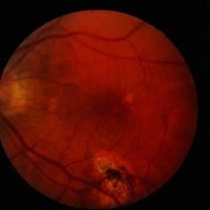
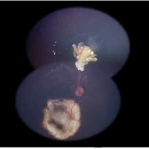 Retinoblastoma Regression
Mar 31 2017 by Andrea Arriola-Lopez, MD MSc
Color photograph shows retinoblastoma regression following treatment.
Photographer: Andrea Elizabeth Arriola-Lopez MD MSc
Imaging device: RetCam II
Condition/keywords: atrophic scar, retinoblastoma
-
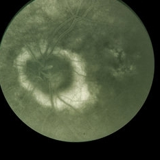
 Presumed Ocular Histoplasmosis Syndrome
Jan 5 2015 by H. Michael Lambert, MD
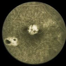
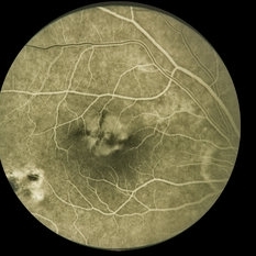
Fluorescein angiogram of OD with temporal atrophic scar and central soft PED with hot cross bun. Presumed Ocular Histoplasmosis Syndrome.
Condition/keywords: presumed ocular histoplasmosis syndrome (POHS)
-
 Presumed Ocular Histoplasmosis Syndrome
Jan 5 2015 by H. Michael Lambert, MD
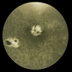
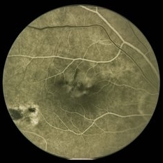
Fluorescein angiogram of OD with temporal atrophic scar and central soft PED with hot cross bun. Presumed Ocular Histoplasmosis Syndrome.
Condition/keywords: presumed ocular histoplasmosis syndrome (POHS)
-
 Presumed Ocular Histoplasmosis Syndrome
Jan 5 2015 by H. Michael Lambert, MD
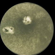
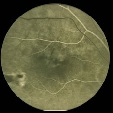
Fluorescein angiogram of OD with temporal atrophic scar and central soft PED with hot cross bun. Presumed Ocular Histoplasmosis Syndrome.
Condition/keywords: presumed ocular histoplasmosis syndrome (POHS)
-
 Presumed Ocular Histoplasmosis Syndrome
Jan 5 2015 by H. Michael Lambert, MD
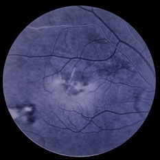
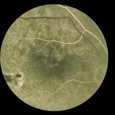
Fluorescein angiogram of OD with temporal atrophic scar and central soft PED with hot cross bun. Presumably Presumed Ocular Histoplasmosis Syndrome.
Condition/keywords: presumed ocular histoplasmosis syndrome (POHS)
-
 Presumed Ocular Histoplasmosis Syndrome
Jan 5 2015 by H. Michael Lambert, MD
No history: fluorescein angiogram of OD with temporal atrophic scar and central soft PED with hot cross bun. Presumed Ocular Histoplasmosis Syndrome.
Condition/keywords: presumed ocular histoplasmosis syndrome (POHS)
-
 Presumed Ocular Histoplasmosis Syndrome
Jan 5 2015 by H. Michael Lambert, MD
Fluorescein angiogram of OD with temporal atrophic scar and central soft PED with hot cross bun. Presumed Ocular Histoplasmosis Syndrome.
Condition/keywords: presumed ocular histoplasmosis syndrome (POHS)
-
 Presumed Ocular Histoplasmosis Syndrome
Jan 5 2015 by H. Michael Lambert, MD
Fluorescein angiogram of OD with temporal atrophic scar and central soft PED with hot cross bun. Presumably Presumed Ocular Histoplasmosis Syndrome.
Condition/keywords: presumed ocular histoplasmosis syndrome (POHS)
-
 Presumed Ocular Histoplasmosis Syndrome
Jan 5 2015 by H. Michael Lambert, MD
Fluorescein angiogram of OD with temporal atrophic scar and central soft PED with hot cross bun. Presumably Presumed Ocular Histoplasmosis Syndrome.
Condition/keywords: presumed ocular histoplasmosis syndrome (POHS)
-
 Presumed Ocular Histoplasmosis Syndrome
Jan 5 2015 by H. Michael Lambert, MD
Fluorescein angiogram of OD with temporal atrophic scar and central soft PED with hot cross bun. Presumably Presumed Ocular Histoplasmosis Syndrome.
Condition/keywords: presumed ocular histoplasmosis syndrome (POHS)
-
 Presumed Ocular Histoplasmosis Syndrome
Jan 5 2015 by H. Michael Lambert, MD
Fluorescein angiogram of OD with temporal atrophic scar and central soft PED with hot cross bun. Presumably Presumed Ocular Histoplasmosis Syndrome.
Condition/keywords: presumed ocular histoplasmosis syndrome (POHS)
-
 Presumed Ocular Histoplasmosis Syndrome
Jan 5 2015 by H. Michael Lambert, MD
Fluorescein angiogram of OD with temporal atrophic scar and central soft PED with hot cross bun. Presumably Presumed Ocular Histoplasmosis Syndrome.
Condition/keywords: presumed ocular histoplasmosis syndrome (POHS)
-
 Presumed Ocular Histoplasmosis Syndrome
Jan 5 2015 by H. Michael Lambert, MD
OD with temporal atrophic scar and central soft PED. Presumably Presumed Ocular Histoplasmosis Syndrome.
Condition/keywords: presumed ocular histoplasmosis syndrome (POHS)
-
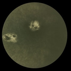
 Presumed Ocular Histoplasmosis Syndrome
Jan 5 2015 by H. Michael Lambert, MD
Presumed Ocular Histoplasmosis Syndrome with atrophic scars and larger inactive lesion inferiorly.
Condition/keywords: presumed ocular histoplasmosis syndrome (POHS)
-
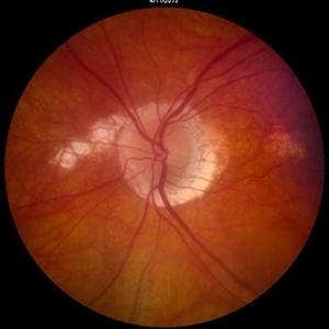
 Choroidal Osteoma
Mar 27 2014 by Jason S. Calhoun
Patient with decreased vision within the past 2-years. VA 20/100 in the right eye. Fundus photo shows atrophic scar temporal to the macula in the right eye.
Photographer: Jason S. Calhoun, Mayo Clinic Jacksonville, Department of Ophthalmology
Imaging device: TOPCON TRC 50-EX
Condition/keywords: choroidal osteoma
-
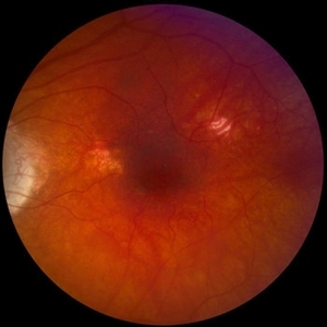
 Treated VKH
May 14 2013 by David Callanan, MD
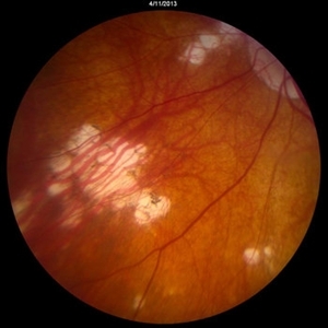
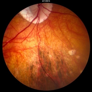
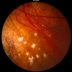
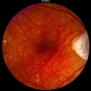
Photos of both eyes of a 66-year-old male with a 4 year history of VKH treated with oral prednisone and mycophenolate. His presenting acuity was hand motions OD and counting fingers at 4 feet OS. Now 20/20-1 OU. Areas of previous inflammatory activity are seen as atrophic scars now.
-
 VKH5
VKH5
May 14 2013 by David Callanan, MD
Photos of both eyes of a 66-year-old male with a 4 year history of VKH treated with oral prednisone and mycophenolate. His presenting acuity was hand motions OD and counting fingers at 4 feet OS. Now 20/20-1 OU. Areas of previous inflammatory activity are seen as atrophic scars now.
-
 VKH4
VKH4
May 14 2013 by David Callanan, MD
Photos of both eyes of a 66-year-old male with a 4 year history of VKH treated with oral prednisone and mycophenolate. His presenting acuity was hand motions OD and counting fingers at 4 feet OS. Now 20/20-1 OU. Areas of previous inflammatory activity are seen as atrophic scars now.
-
 VKH3
VKH3
May 14 2013 by David Callanan, MD
Photos of both eyes of a 66-year-old male with a 4 year history of VKH treated with oral prednisone and mycophenolate. His presenting acuity was hand motions OD and counting fingers at 4 feet OS. Now 20/20-1 OU. Areas of previous inflammatory activity are seen as atrophic scars now.
-
 VKH2
VKH2
May 14 2013 by David Callanan, MD
Photos of both eyes of a 66-year-old male with a 4 year history of VKH treated with oral prednisone and mycophenolate. His presenting acuity was hand motions OD and counting fingers at 4 feet OS. Now 20/20-1 OU. Areas of previous inflammatory activity are seen as atrophic scars now.
-
 VKH1
VKH1
May 14 2013 by David Callanan, MD
Photos of both eyes of a 66-year-old male with a 4 year history of VKH treated with oral prednisone and mycophenolate. His presenting acuity was hand motions OD and counting fingers at 4 feet OS. Now 20/20-1 OU. Areas of previous inflammatory activity are seen as atrophic scars now.
-
---thumb.jpg/image-square;max$300,300.ImageHandler) Age Related Macular Degeneration - Geographic Atrophy
Age Related Macular Degeneration - Geographic Atrophy
May 3 2013 by Suber S. Huang, MD, MBA, FASRS
Geographic Atrophy.
Imaging device: Retina Diseases Imaging Analysis Reading Center
Condition/keywords: advanced geographic atrophy, atrophic scar, atrophic spot, geographic atrophy, macula lesion, pigment epithelial atrophy
-
---thumb.jpg/image-square;max$300,300.ImageHandler) Age Related Macular Degeneration - Geographic Atrophy
Age Related Macular Degeneration - Geographic Atrophy
May 3 2013 by Suber S. Huang, MD, MBA, FASRS
Geographic Atrophy.
Imaging device: Retina Diseases Imaging Reading Center
Condition/keywords: advanced geographic atrophy, atrophic scar, atrophic spot, geographic atrophy, macula lesion, pigment epithelial atrophy, red-free, window defect
-
---thumb.jpg/image-square;max$300,300.ImageHandler) Age Related Macular Degeneration - Geographic Atrophy
Age Related Macular Degeneration - Geographic Atrophy
May 3 2013 by Suber S. Huang, MD, MBA, FASRS
Geographic Atrophy.
Imaging device: Retina Diseases Imaging Analysis Reading Center
Condition/keywords: advanced geographic atrophy, atrophic scar, atrophic spot, geographic atrophy, macula lesion, pigment epithelial atrophy
 Loading…