Search results (3 results)
-
 Tributary vein occlusion
Sep 26 2023 by Ben Serar
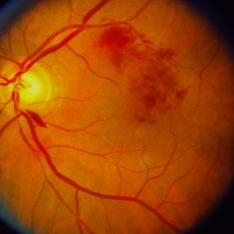
Fundus photograph of LE showing retinal haemorrhages along the superotemporal arcade , with splinter haemorrhage near the disc, in a case of tributary vein occlusion.
Condition/keywords: Tributary vein occlusion
-
 Myelinated Nerve Fibres in left eye with old tributary vein occlusion in left eye
Jul 18 2023 by Harsh Vardhan Singh, MS
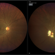
55 year female with left eye amblyopia & high myopia with MNF and Right eye showed signs of old macular branch retinal vein occlusion
Photographer: Harsh Vardhan Singh, AIIMS, Guwahati
Imaging device: Zeiss Clarus 700
Condition/keywords: BRVO, macular branch retinal vein occlusion (BRVO), Medullated Nerve fibres, MNF, Myelinated Nerve Fibres, TRVO
-
 Peri-papillary Vascular Loop
Jun 2 2020 by Dhaivat Shah
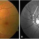
Peri-papillary vascular loops (PVL) are rare congenital vascular malformations, which are usually detected as accidental finding during routine fundus examination. They can often be confused with tributary vein occlusion or racemose hemangioma. Although benign and asymptomatic, they can be rarely associated with vitreous hemorrhage and arterial occlusion. We herein present a case of a 60-year-old hypertensive male, who was diagnosed elsewhere to have a tributary vein occlusion and was referred to us. FFA was advised to rule out neovascularization, surrounding capillary non perfusion and mass lesion (hemangioma). On FFA, the arterial loop showed a slightly delayed filling (3-5 seconds) as compared to the other arterial vessels and the original vessel appeared to be a branch arising from central retinal artery. The choroidal filling was delayed in the area supplied by the loop. A cilioretinal artery was also noted. The patient was diagnosed to have a Peri-papillary vascular arterial loop (PVL), likely to be congenital in origin. The patient was reassured and was advised yearly follow up. These loops are usually accidental findings discovered during routine fundus examination. Since these vessels are looped and tortuous, they exhibit a slower and laminar blood flow, which make them more prone for arterial occlusions. The vitreous in this area tends to be adherently attached, so during PVD induction, it is likely to cause a tear and hemorrhage leading to vitreous hemorrhage. Until and unless there is a break, this hemorrhage tends to resolve on its own and does not warrant treatment. If there is an evident break, it can be dealt with laser barrage.
Photographer: Choithram Netralaya
Condition/keywords: congenital prepapillary vascular loop
 Loading…