Search results (177 results)
-
 Stars of Stargardt
Aug 4 2025 by Malvika Singh
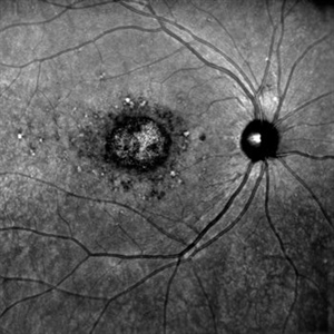
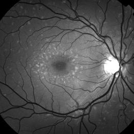
Infrared fundus photograph of a 22 year old female with Stargardt's disease.
Photographer: Dr Malvika Singh, Retina Foundation, Ahmedabad, India
Imaging device: Mirante SLO/OCT
Condition/keywords: infrared image, Stargardt disease
-
 Stargardt's Disease (Extensive)
Jul 16 2025 by Shivankar Sen, MS, FVRS
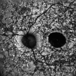
15-year-old female with complaints of defective vision for 6 years with best corrected visual acuity of 6/60; N36 in both eyes was found to have dystrophic macula with extensive spread out pigmentary bony spicules; On Confocal blue autofluorescence shows central hypo-autofluorescence and a heterogenous pisciform background with OCT showing extensive outer retinal layer disruption with genetic report confirming ABCA4 mutation and giving us definite diagnosis of Stargardt's Disease.
Photographer: Dr. N. Haindavi
Imaging device: Heidelberg Spectralis HRA+OCT
Condition/keywords: Blue autofluroscence, Stargardts Disease
-
 Hereditary Macular Dystrophy with CNVM
Apr 4 2025 by Tejaswita Verma
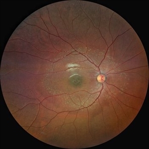
Fundus photo of a 46 y/o male with 6/36 vision, Stargardt's disease with CNVM
Photographer: DR. TEJASWITA VERMA
Imaging device: MIRANTE
Condition/keywords: CNVM, hereditary macular dystrophy, HMD
-
 Stargardt's Disease
Oct 23 2024 by Virginia Gebhart
62 year old female with bullseye RPE changes and flecks, mottled FAF, and silent choroid on FA consistent with late onset Stargardt's Disease. Pt is asymptomatic with 20/20 vision OU at this time
Photographer: Virginia Gebhart, Retina Consultants of Carolina
Imaging device: Optos California
Condition/keywords: Stargardt disease, Stargardts Disease
-
 Fundus Flavimaculatus Fundus Autofluorescence Imaging
Sep 25 2024 by Keshavi Shah
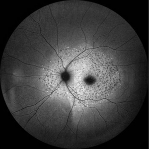
FAF imaging of a 37 year old male patient with Stargardt's Disease of adult onset ( Fundus Flavimaculatus) presenting with dimunition of night vision and dyschromatopsia demonstrating areas of hypo-auto fluorescence (representing RPE/ Photo-receptor atrophy) and hyper-autofluorescence(representing excessive lipo-fuschin accumulation in the RPE cells) with peri-papillary sparing, typical of ABCA-4 related disorders.
Photographer: Simran
Imaging device: Optos Daytona
Condition/keywords: fundus autofluorescence (FAF), fundus flavimaculatus
-
 Fundus Flavimaculatus Fundus Autofluorescence Imaging
Sep 25 2024 by Keshavi Shah
FAF imaging of a 37 year old male patient with Stargardt's Disease of adult onset ( Fundus Flavimaculatus) presenting with dimunition of night vision and dyschromatopsia demonstrating areas of hypo-auto fluorescence (representing RPE/ Photo-receptor atrophy) and hyper-autofluorescence(representing excessive lipo-fuschin accumulation in the RPE cells) with peri-papillary sparing, typical of ABCA-4 related disorders.
Photographer: Simran
Imaging device: Nikon Optos Daytona
Condition/keywords: fundus autofluorescence (FAF), fundus flavimaculatus
-
 B-FAF in Stargardt's Disease
Jul 4 2024 by Tejaswita Verma
Blue fundus autofluorescence showing hypoautofluorescence picture of a 28 year old male with 6/60 vision in BE in a case of Stargardt's disease.
Photographer: DR. TEJASWITA VERMA
Imaging device: MIRANTE
Condition/keywords: fundus autofluorescence (FAF), hereditary macular dystrophy, Stargardt disease
-
 Stargardt's Disease
Apr 20 2024 by Tejaswita Verma
Fundus autofluorescence image of the left eye of a 39 year old male showing hypoautofluorescence in a case of Stargardt's disease.
Photographer: DR. TEJASWITA VERMA
Imaging device: MIRANTE
Condition/keywords: fundus autofluorescence (FAF), hereditary macular dystrophy, hypoautofluorescence, Stargardt disease
-
 Stargardt's Disease
Apr 20 2024 by Tejaswita Verma
Fundus autofluorescence image of the right eye of a 39 year old male showing hypoautofluorescence in a case of Stargardt's disease.
Photographer: DR. TEJASWITA VERMA
Imaging device: MIRANTE
Condition/keywords: fundus autofluorescence (FAF), hereditary macular dystrophy, hypoautofluorescence, Stargardt disease
-
 Stargardt's Disease
May 5 2023 by Virginia Gebhart
51-year-old male with bilateral central retinal dystrophy consistent with Stargardt disease. No significant progression of central atrophy, and VA has remained stable at 20/150 since 2012
Photographer: Virginia Gebhart, Retina Consultants of Carolina
Imaging device: Topcon TRC 50DX
Condition/keywords: Stargardt disease
-
 Macular Dystrophy
Aug 31 2022 by Jeffrey Barker
Genetic Macular Dystrophy, most likely not Stargardt's Disease.
Photographer: Jeffrey P. Barker, B.S.
Imaging device: Topcon TRC-50EX
Condition/keywords: macular dystrophy, Stargardt disease
-
 Stargardt's Disease
Aug 18 2021 by Priyanka Raj, MBBS, MS
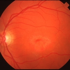
Fundus photograph of a 24 year-old male with Stargardt's disease
Photographer: Priyanka Raj, Prakash Netra Kendr, Lucknow, India
Imaging device: Zeiss Clarus 500
Condition/keywords: fundus flavimaculatus, heredomacular degeneration, Stargardt disease
-
 Stargardt's disease
Aug 18 2021 by Priyanka Raj, MBBS, MS
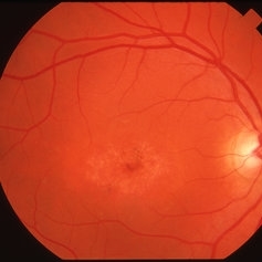
Fundus photograph of a 24 year-old male with Stargardt's disease
Photographer: Priyanka Raj, Prakash Netra Kendr, Lucknow, India
Imaging device: Zeiss Clarus 500
Condition/keywords: fundus flavimaculatus, Stargardt disease
-
 Stargardt's disease
Aug 18 2021 by Priyanka Raj, MBBS, MS
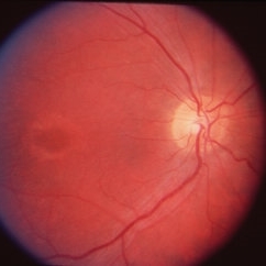
Fundus photograph of a 24 year-old male with Stargardt's disease
Photographer: Priyanka Raj, Prakash Netra Kendr, Lucknow, India
Imaging device: Zeiss Clarus 500
Condition/keywords: fundus flavimaculatus, heredomacular degeneration, Stargardt disease
-
 Stargardt's disease
Aug 18 2021 by Priyanka Raj, MBBS, MS
Fundus photograph of a 24 year-old male with Stargardt's disease.
Photographer: Priyanka Raj, Prakash Netra Kendr, Lucknow, India
Imaging device: Zeiss Clarus 500
Condition/keywords: fundus flavimaculatus, heredomacular degeneration, Stargardt disease
-
 Stargardt's Disease
May 27 2020 by Jamin S. Brown, MD
Fundus autofluorescence image of 46-year-old male with Stargardt's Disease.
Photographer: Jeffrey Barker, Retina-Vitreous Surgeons of CNY
Condition/keywords: Stargardt disease
-
 Stargardt's Disease
Apr 8 2019 by Gary R. Cook, MD, FACS
38-year-old white male with atrophic macular lesion OS secondary to Stargardt's disease; V.A. = 20/200
Imaging device: Topcon VT-50
Condition/keywords: atrophic central lesion, Stargardt disease
-
 Stargardt's Disease
Apr 8 2019 by Gary R. Cook, MD, FACS
38-year-old white male with typical atrophic macular lesion OD secondary to Stargardt's disease; V.A. = 20/200
Imaging device: Topcon VT-50
Condition/keywords: atrophic central lesion, Stargardt disease
-
 Stargardt's Disease
Apr 3 2019 by Gary R. Cook, MD, FACS
19-year-old white male with atrophic maculopathy OS secondary to Stargardt's disease; V.A. = 20/200.
Condition/keywords: atrophic central lesion, Stargardt disease
-
 Stargardt's Disease
Apr 3 2019 by Gary R. Cook, MD, FACS
19-year-old white male with atrophic maculopathy OD secondary to Stargardt's disease; V.A. = 200
Condition/keywords: atrophic central lesion, Stargardt disease
-
 Stargardt's Disease
Dec 17 2018 by Deepak Bhojwani, MS
Fundus image and autofluorescence image of left eye a 26- year-old male with large area of RPE atrophy over macular region surrounded by pisciform flecks. Note the flecks are better appreciated on autofluorescence images (classic hypofluorescent pisciform lesions).
Photographer: Dr. Deepak Bhojwani
Imaging device: AUTO FLOROSCENCE IMAGING
Condition/keywords: autofluorescence imaging, macular dystrophy, Stargardt disease
-
 Stargardt's Disease
Oct 4 2018 by Aditya S Kelkar, MS, FRCS, FASRS,FRCOphth
Auto-fluorescence image of a 19-year-old male showing flecks of both increased and decreased autofluorescence and reduced central macular autofluorescence surrounded by an increased signal.
Photographer: Dr. Aanchal Agarwal
Condition/keywords: Stargardt disease
-
 Stargardt's Disease - Dark Choroid
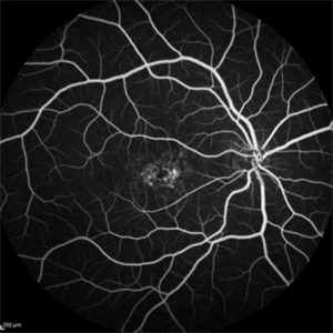
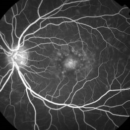
Dec 1 2016 by Courtney Crawford, MD, FACS
24-year-old male with progressive decreased vision to level of 20/200 OU.
Photographer: Kristen Dunn, North Texas Retina Consultants
Condition/keywords: choroid, Stargardt disease
-
 Stargardt's disease
Jun 11 2016 by John S. King, MD
RF
Condition/keywords: bull's eye maculopathy, fleck dystrophy, Stargardt disease
-
 Stargardt's Disease
Jun 11 2016 by John S. King, MD
Dark choroid.
Condition/keywords: bull's eye maculopathy, fleck dystrophy, Stargardt disease
 Loading…