Search results (1816 results)
-
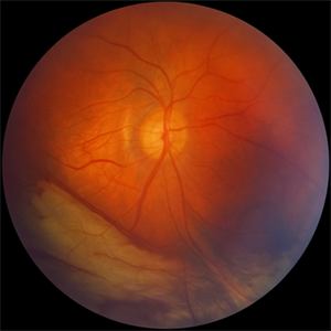
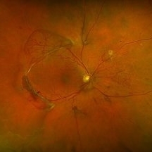
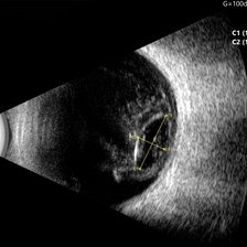
Color Rhegmatogenous Retinal Detachment
Aug 13 2025 by Gustavo Uriel Fonseca Aguirre
Color Doppler ultrasound of Rhegmatogenous Retinal Detachment.
Condition/keywords: color doppler, Rhegmatogenous retinal detachment
-
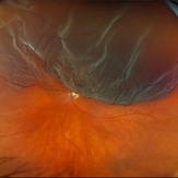
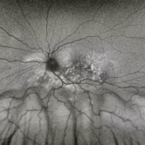
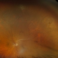
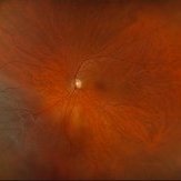
 Post Vitreoretinal Surgery
Aug 13 2025 by Debarun Sharma
Fundus photograph of the same patient after successful repair of retinal detachment with choroidal drainage by pars plana vitrectomy with silicone oil tamponade.
Photographer: Debarun Sharma, Sri Sankardeva Nethralaya, Guwahati
Imaging device: Optos
Condition/keywords: vitreoretinal surgery
-
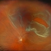
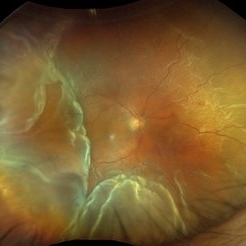
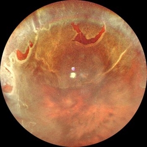
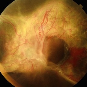
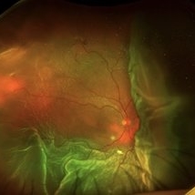 Retinal Detachment with Large Choroidal Detachment
Aug 13 2025 by Debarun Sharma
Fundus photograph of a 56 year old man with rhegmatogenous retinal detachment with a large nasal choroidal detachment.
Photographer: Debarun Sharma, Sri Sankardeva Nethralaya, Guwahati
Imaging device: Optos
Condition/keywords: choroidal detachment, retinal detachment
-
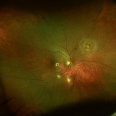
 Macular Hole Due to Proliferative Diabetic Retinopathy
Aug 13 2025 by Ricardo Leitão Guerra
A macular hole formation after anti-VEGF injection prior to vitrectomy for tractional retinal detachment in a patient presenting proliferative diabetic retinopathy.
Photographer: Ricardo Leitão Guerra
Imaging device: ZEISS CLARUS 700
Condition/keywords: macular hole, proliferative diabetic retinopathy (PDR)
-
 Amelanotic Melanoma
Aug 12 2025 by César Adrián Gómez Valdivia, MD
This case highlights an amelanotic melanoma, an atypical presentation of a choroidal melanoma lacking the characteristic pigmentation. These lesions can easily be mistaken for choroidal hemangiomas, metastases, or inflammatory masses. Clinically, the lesion appears as a dome-shaped, yellowish subretinal mass, often associated with subretinal fluid, lipofuscin deposition, or retinal detachment. The absence of pigment can delay diagnosis, making multimodal imaging essential. Diagnostic tools: • B-scan ultrasound: low to medium internal reflectivity • OCT: overlying subretinal fluid and RPE elevation • FAF: orange pigment and RPE disruption • ICG/FA: variable, often hypofluorescent core Important: Prompt referral to ocular oncology is critical for management and prognosis.
Photographer: @eyemissu2
Imaging device: TOPCON TRC-50DX
Condition/keywords: amelanotic melanoma
-
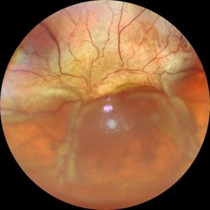
 Retinal Detachment
Aug 12 2025 by Kimberly Wakester
Optomap RGB of a 63-year-old man with a bullous overhanging Retinal Detachment with superonasal tuft-associated tear, macula detached in the left eye. Surgery was recommended. Patient is to continue follow up care post operatively.
Photographer: Kimberly Wakester, COA, OCT-C, Retina Consultants of Carolina
Imaging device: Optos California
Condition/keywords: bullous retinal detachment, left eye, Mac off
-
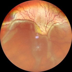
 Retinal Detachment with Multiple Breaks
Aug 12 2025 by Kimberly Wakester
Optomap RGB of a 59-year-old man with a retinal detachment with multiple breaks in the left eye. Surgery was recommended. Patient is to continue follow up care post operatively.
Photographer: Kimberly Wakester, COA, OCT-C, Retina Consultants of Carolina
Imaging device: Optos California
Condition/keywords: lattice degeneration, left eye, Retinal Detachment with Multiple Breaks
-
 Retinal Dialysis Associated Detachment
Aug 7 2025 by Elaine Michele Binkley, MD
Retinal detachment associated with large retinal dialysis.
Photographer: Alexandra Copple, University of Iowa, Iowa City, IA
Condition/keywords: Retinal dialysis
-
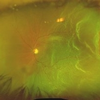
 Exudative Retinal Detachment
Aug 6 2025 by Aditya S Kelkar, MS, FRCS, FASRS,FRCOphth
Fundus auto-fluorescence of a 41 year old female depicting retinal pigment epitheliopathy and exudative retinal detachment in case of ocular metastasis secondary to breast carcinoma.
Photographer: Dr.Rabia Naaz, National Institute of ophthalmology, Pune
Imaging device: OPTOS DAYTONA
Condition/keywords: Exudative retinal detachment, Retinal pigment epitheliopathy
-
 Total Retinal Detachment
Aug 6 2025 by Korey Starkey
59 year-old patient presents with total retinal detachment at first visit in OD. Recommending prompt surgical intervention.
Photographer: Korey Starkey
Imaging device: Optos
Condition/keywords: color fundus photograph, Optos, retinal detachment, total retinal detachment
-
 Horseshoe Retinal Tear
Aug 6 2025 by Korey Starkey
80 year-old patient presented with HSRT without detachment in the left eye and macula-off detachment in the right eye. Scheduled patient for prompt surgical repair OD and same day laser retinopexy OS to reduce risk of retinal detachment.
Photographer: Korey Starkey
Imaging device: Optos
Condition/keywords: color fundus photograph, fundus photography, horseshoe tear, Optos
-
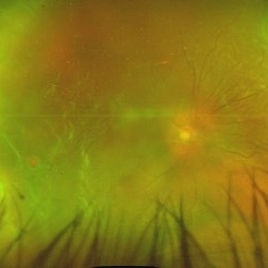
 Traction in Progression
Aug 6 2025 by Claudio Brancato, MD
This image captures stage 4A Retinopathy of Prematurity, showing partial retinal detachment sparing the macula. The elevated retina and fibrous ridge indicate tractional forces secondary to extraretinal neovascularization. A striking representation of disease evolution, poised between reversibility and vision loss.
Photographer: Gregorio Lo Giudice, ARNAS Civico Hospital, Palermo, Italy
Imaging device: RETCAM 3 (enhanced via IA)
Condition/keywords: retinopathy of prematurity
-
 Retinal Detachment Secondary to Large Temporal Tear
Aug 4 2025 by Anjana Mirajkar, MS Ophthalmology
Fundus photograph of a 55 year old male with a retinal detachment with macula off with a large temporal tear.
Photographer: Dr. Anjana Mirajkar- HV desai eye hospital ,Pune
Imaging device: Optos
Condition/keywords: Retinal Detachment, retinal tear
-
 Unstable PDR s/p Laser
Aug 4 2025 by Anjana Mirajkar, MS Ophthalmology
Fundus photograph of a 60 year old male with an unstable PDR showing traction at the posterior pole with sub hyaloid hemorrhage. Peripheral PRP marks can be seen.
Photographer: Dr. Anjana Mirajkar- HV Desai eye hospital ,Pune
Imaging device: Optos
Condition/keywords: pan-retinal photocoagulation (PRP), proliferative diabetic retinopathy (PDR), subhyaloid hemorrhage, tractional retinal detachment
-
 Retinal Detachment
Aug 4 2025 by Anjana Mirajkar, MS Ophthalmology
Fundus photograph of a 40 year old male with a total retinal detachment with macula off with a HST at 1 o clock and a break at mid periphery at 8 o clock.
Photographer: Dr. Anjana Mirajkar- HV Desai eye hospital ,Pune
Imaging device: optos
Condition/keywords: retinal detachment, retinal holes
-
 Chronic RD with Retinal Dialysis
Jul 23 2025 by Virginia Gebhart
64 year old female with chronic retinal detachment from head trauma 41 years ago. Peripheral scarring from 6:00 to 11:00 with area of subretinal fluid inferotemporally, well demarcated with subretinal bands. Retinal dialysis inferotemporal from 7:00 to 9:00. No surgical repair needed or recommended at this time.
Photographer: Virginia Gebhart, Retina Consultants of Carolina
Imaging device: Optos California
Condition/keywords: chronic retinal detachment, demarcation, RD, Retinal Detachment, retinal dialysis, subretinal bands
-
 Retinal detachment with Single Break
Jul 18 2025 by Kimberly Wakester
Optomap RGB of a 62-year-old man with a retinal detachment with a single break in the left eye. Patient has a previously treated HSRT in the left eye. Surgery was recommended. Patient is to continue follow up care post operatively.
Photographer: Kimberly Wakester, COA, OCT-C
Imaging device: Optos California
Condition/keywords: RD, retinal tear
-
 Retinal Detachment with Multiple Horse Shoe Shaped Tears
Jul 14 2025 by Malvika Singh
Fundus photograph of a 46 year old showing a retinal detachment with multiple peripheral horse show shaped tears.
Photographer: Dr Malvika Singh, Retina Foundation, Ahmedabad, India
Imaging device: Mirante SLO/OCT
Condition/keywords: horseshoe tear, retinal detachment
-
 Exudative Retinal Detachment
Jul 12 2025 by Akansha Sharma
Color fundus photograph of a 33 year old male with exudative retinal detachment with a small axial length.
Photographer: DR. AKANSHA SHARMA
Condition/keywords: exudative detachment
-
 Exudative Retinal Detachment
Jul 12 2025 by Akansha Sharma
Color fundus photograph of a 33 year old male with exudative retinal detachment with a small axial length.
Photographer: DR. AKANSHA SHARMA
Condition/keywords: exudative detachment
-
 Proliferative Diabetic Retinopathy
Jul 9 2025 by Jeffrey Barker
57 year old male presents with Proliferative Diabetic Retinopathy and Tractional Retina detachment.
Photographer: Jeffrey P. Barker, B.S. Retina Vitreous Surgeons of CNY
Condition/keywords: Diabetes, proliferative diabetic retinopathy (PDR), Traction retinal detachment
-
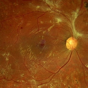
 Annular Tractional Retinal Detachment
Jul 5 2025 by César Adrián Gómez Valdivia, MD
Fundus photograph of an 66 YO female patient diagnosed with advanced proliferative diabetic retinopathy.
Photographer: @eyemissu2
Imaging device: TOPCON TRC-50DX
Condition/keywords: tractional retinal detachment
-
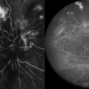 Combined Occlusion with TRD
Jun 30 2025 by Shivankar Sen, MS, FVRS
Posterior Pole and Ultra-wide field Fluorescein angiogram of a 79 yr. old one eyed male revealing arterial occlusion, grossly non-perfused peripheral retina with neovascularisation elsewhere and significant tractions at the posterior pole.
Photographer: Gayathri M S, Dr. Shivankar Sen MD
Imaging device: Heidelberg Spectralis HRA+OCT
Condition/keywords: arterial occlusion, Traction retinal detachment, Vein Occlusion
-
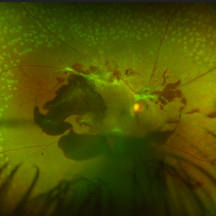
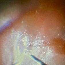 Love Through the Lens of Retinal Detachment
Jun 27 2025 by Claudio Brancato, MD
The image depicts a case of rhegmatogenous retinal detachment where the vitreous was extremely adherent to the retina. The primary surgeon was performing membrane peeling using a surgical loop, while the assisting surgeon was captivated by the intricate procedure. In a moment of affectionate dedication, the primary surgeon carefully peeled the membrane to form a heart shape, symbolizing both his passion for surgery and perhaps a personal gesture towards the assisting surgeon. This delicate and precise maneuver highlights the complexity and artistry involved in vitreoretinal surgery, showcasing the blend of technical skill and emotional expression within the operating room.
Photographer: Claudio Brancato, ARNAS CIVICO Hospital, Palermo, Italy
Imaging device: Zeiss Artevo 800
Condition/keywords: finesse, peeling, proliferative vitreoretinopathy (PVR), Retina detachment
-
 Ocular B-scan Ultrasound (Longitudinal Scan M6, gain 100 dB)
Jun 26 2025 by Hector Gabriel Moreno Solano, MD, MHA
B-scan ultrasound was performed in longitudinal section M6 with a gain of 100 dB. A hyperechoic structure with posterior acoustic shadowing is observed, consistent with lens luxation and condensed vitreous bands adjacent to the lens. The dislocated lens measures approximately 9.54 mm x 4.62 mm. The study was conducted following blunt ocular trauma caused by a golf ball. The remaining vitreous cavity appears anechoic, with no evidence of retinal detachment or other structural abnormalities in this section.
Photographer: Hector Gabriel Moreno Solano, Instituto Mexicano de Oftalmología “IMO I.A.P”
Imaging device: Quantel Medical
Condition/keywords: B scan ultrasound, lens luxation, ocular trauma
 Loading…