Search results (521 results)
-
 Nevus to Melanoma in 1 Year
Dec 12 2025 by Virginia Gebhart
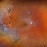
83 year old male presents with partially amelanotic choroidal nevus, which has been followed for the last 10 years. On exam there is a new area of orange pigment, and change in diameter compared to photos from 2024. Ultrasound shows small increase in height, medium to low internal reflectivity, and vascularity. Pt is asymptomatic. Due to personal matters pt wishes to reassess in few months and schedule treatment.
Photographer: Virginia Gebhart, Retina Consultants of Carolina
Imaging device: Optos California
Condition/keywords: choroidal melanoma, choroidal nevus
-
 CHRPE
CHRPE
Dec 11 2025 by Virginia Gebhart
48 year old female referred for pigmented lesion. Exam and photos consistent with well circumscribed CHRPE with lacunae. Patient previously unaware. Observation recommended.
Photographer: Virginia Gebhart, Retina Consultants of Carolina
Imaging device: Optos California
Condition/keywords: CHRPE, congenital hypertrophy of the retinal pigment epithelium (CHRPE)
-
 Retinitis Pigmentosa
Dec 9 2025 by Kimberly Wakester
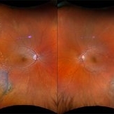
Optomap RGB and AF of an 78-year-old woman with Retinitis Pigmentosa. Patient is to continue follow up care every 6 months to monitor progression.
Photographer: Kimberly Wakester, COA, OCT-C
Imaging device: Optos California
Condition/keywords: retinitis pigmentosa
-
 Retinal Detachment with HSRT
Dec 9 2025 by Kimberly Wakester
Optomap RGB of an 43-year-old woman with a retinal detachment with HSRT in the right eye. Surgery was recommended. Patient is to continue follow up care post operatively.
Photographer: Kimberly Wakester, COA, OCT-C
Imaging device: Optos California
Condition/keywords: retinal detachment with HSRT
-
 Retinal Detachment with PVR
Dec 9 2025 by Kimberly Wakester
Optomap RGB of an 69-year-old man with a retinal detachment with PVR formation inferotemporal in the right eye s/p RD repair. Surgery was recommended. Patient is to continue follow up care post operatively.
Photographer: Kimberly Wakester, COA, OCT-C
Imaging device: Optos California
Condition/keywords: gas bubble, retinal detachment with PVR
-
 Chronic RD with Retinoschisis
Dec 9 2025 by Kimberly Wakester
Optomap RGB of an 67-year-old woman with a chronic retinal detachment with retinoschisis in the left eye. Surgical intervention vs observation was discussed with patient. Recommended close observation at this time to monitor the progression of the SRF prior to surgical intervention. Patient will return in 6-8 weeks to repeat dilated exam and imaging.
Photographer: Kimberly Wakester, COA, OCT-C
Imaging device: Optos California
Condition/keywords: Chronic RD, retinoschisis
-
 Best Disease
Dec 9 2025 by Kimberly Wakester
Optomap RBG and AF photograph of an 65-year-old man with Best disease in the left eye. The hypopigmented lesions appear stable on clinical exam and fundus photos compared to previous images. Patient is to continue yearly follow up care with dilated exam and repeat imaging.
Photographer: Kimberly Wakester, COA, OCT-C
Imaging device: Optos California
Condition/keywords: Best Disease, Dystrophies of the Retinal Pigment Epithelium
-
 Rhegmatogenous Retinal Detachment
Nov 27 2025 by Jacob Adrián Ruíz García
Ultra–widefield fundus image demonstrates an extensive rhegmatogenous retinal detachment (RRD) with several large, irregular retinal tears are visible in the temporal retina, with associated surrounding subretinal fluid. The detachment appears bullous, with fluid extending widely across the mid-periphery and involving much of the posterior pole.
Photographer: Jacob Adrián Ruíz Garcia, Fundación Hospital Nuestra Señora de la Luz I.A.P, México City
Imaging device: Optos California
Condition/keywords: horseshoe tear, Rhegmatogenous retinal detachment, tear
-
 Traction Detachment of Retina
Nov 14 2025 by Virginia Gebhart
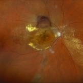
50 year old female with proliferative diabetic retinopathy, large ridge of traction temporally, and significant band of fibrosis. Subretinal fluid throughout the macula. Due to traction, surgical repair not recommended at this time as it could worsen condition. Will observe closely. BCVA CF @ 1 ft.
Photographer: Virginia Gebhart, Retina Consultants of Carolina
Imaging device: Optos California
Condition/keywords: fibrosis, proliferative diabetic retinopathy (PDR), traction detachment, Traction retinal detachment
-
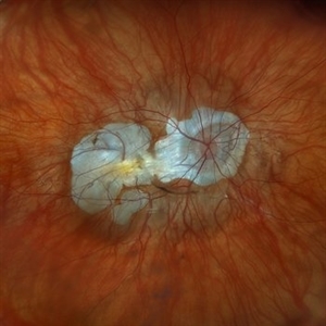
 Bullous Retinal Detachment
Nov 13 2025 by Virginia Gebhart
42 year old female referred for vision loss x 4-5 days. Bullous retinal detachment from 8:00 to 3:00 with retinal tear at 11:00. Macula is detached. Vision is LP, IOP of 3. Pt is scheduled for GFE and possible scleral buckle.
Photographer: Virginia Gebhart, Retina Consultants of Carolina
Imaging device: Optos California
Condition/keywords: bullous retinal detachment, retinal detachment, retinal detachment of the macula, retinal tear with detachment
-
 Giant Nevus of the Macula
Nov 5 2025 by Virginia Gebhart
68 year old female with stable large pigmented lesion throughout the whole posterior pole, extending beyond the superior and inferior arcades. Pt has been aware of nevus for 45 years. Lesion has been stable since initial photos in 2013. Will continue to observe. Vision CF
Photographer: Virginia Gebhart, Retina Consultants of Carolina
Imaging device: Optos California
Condition/keywords: Choroidal nevus, pigmented lesion, pigmented nevus
-
 Chorioretinitis
Oct 30 2025 by Kimberly Wakester
Optomap RGB of an 37-year-old-woman with stable Chorioretinitis in the right eye. Patient is to return in 6 months for dilated exam and repeat diagnostic testing.
Photographer: Kimberly Wakester, COA, OCT-C
Imaging device: Optos California
Condition/keywords: chorioretinitis
-
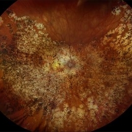
 Pigmentary Retinal Dystrophy
Oct 30 2025 by Kimberly Wakester
Optomap RGB of an 77-year-old-woman with Pigmentary Retinal Dystrophy in the left eye. Patient is to continue follow up care yearly with dilated exam and diagnostic testing.
Photographer: Kimberly Wakester, COA, OCT-C
Imaging device: Optos California
Condition/keywords: bone spicules, Pigmentary Retinal Dystrophy
-
 Elmiron Toxicity
Oct 30 2025 by Kimberly Wakester
Optomap RGB and Optomap AF of an 52-year-old woman with Elmiron toxicity from the use of Elmiron for 19 years. Patient is to continue follow up care every 3-4 months with repeat exam and testing to continue monitoring the progression of the degeneration.
Photographer: Kimberly Wakester, COA, OCT-C
Imaging device: Optos California
Condition/keywords: Elmiron Toxicity
-
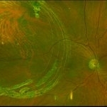
 Vasoproliferative Tumor
Oct 27 2025 by Virginia Gebhart
34 year old male with retinal vasoproliferative tumor with traction detachment temporally and striae in the macula. Recommend vitrectomy prior to cryotherapy to release vitreous from retina. BCVA 20/30
Photographer: Virginia Gebhart, Retina Consultants of Carolina
Imaging device: Optos California
Condition/keywords: traction retinoschisis, tractional retinal detachment, Vasoproliferative Tumor
-
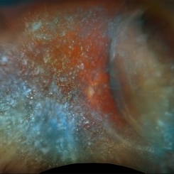 Choroidal and Near Total RD, Severe Asteroid Hyalosis, Treated Melanoma
Oct 22 2025 by Virginia Gebhart
78 year old male with sudden decrease in vision. Poor view due significant asteroid hyalosis. Bscan showed large nasal choroidal and near total retinal detachments, attached temporally. No obvious break found. Regressed tumor inferiorly s/p brachytherapy in April 2023. BCVA 20/320, IOP of 03. Pt schedule for primary PPV and possible SB placement vs. GFE
Photographer: Virginia Gebhart, Retina Consultants of Carolina
Imaging device: Optos California
Condition/keywords: asteroid hyalosis, brachytherapy, choroidal detachment, choroidal melanoma, melanoma, RD, retinal detachment, sub-total retinal detachment
-
 Multifocal Choroiditis with Panuveitis
Oct 16 2025 by Virginia Gebhart
39 year old female diagnosed with MCP in 2009. Extensive RPE changes and hypertrophy, arterial attenuation and pale nerve. Currently no active inflammation.
Photographer: Virginia Gebhart, Retina Consultants of Carolina
Imaging device: Optos California
Condition/keywords: corticosteroid-induced glaucoma, hypertrophy, multifocal chorioretinitis (MCP), PALE DISC
-
 Asteroid Hyalosis
Oct 8 2025 by Virginia Gebhart
49 year old female with asteroid hyalosis. Pt only notices when in bright light.
Photographer: Virginia Gebhart, Retina Consultants of Carolina
Imaging device: Optos California
Condition/keywords: asteroid hyalosis
-
 RD Repair 12 Years Later
Oct 7 2025 by James B. Soque, CRA, OCT-C, COA, FOPS
https://imagebank.asrs.org/file/15894/retinal-detachment-right-eye-optomap See the previous submission above using an Optos TX 2000. Now, 12 years after his RD OD surgery, he remains attached. Comparatively one of the most amazing cases I've been privileged to be part of. This RD Repair from 2014 still going strong! Camera: Optos California
Photographer: James B, Soque, CRA, OCT-C, COA, FOPS, VRC of New York, Shirley, NY
Imaging device: Optos California R/G/B
Condition/keywords: Afx, GasX, Optomap, OPTOS CALIFORNIA, PPV OD, RD Repair 2014, UWF FC
-
 Retinal Detachment with Multiple Breaks
Oct 3 2025 by Kimberly Wakester
Optomap RGB of an 60-year-old man with a retinal detachment with multiple breaks in the left eye. Surgery was recommended. Patient is to continue follow up care post operatively.
Photographer: Kimberly Wakester, COA, OCT-C
Imaging device: Optos California
Condition/keywords: Mac off, Retinal Detachment with multiple breaks
-
 Dry AMD, Advanced Atrophic with Subfoveal Involvement
Oct 3 2025 by Kimberly Wakester
Optomap RGB of an 76-year-old woman with Dry AMD, Advanced Atrophic with Subfoveal Involvement in the right eye. Longstanding, more likely due to myopic/choroidal atrophy. End-stage disease, would not recommend aggressive intervention. Patient is to continue follow up care and repeat OCT/imaging as directed per doctor. Just a fun mention about the image, if you look at the optic nerve and the vessels it appears to look like an eye.
Photographer: Kimberly Wakester, COA, OCT-C
Imaging device: Optos California
Condition/keywords: Advanced Atrophic with Subfoveal Involvement, dry age-related macular degeneration (dry AMD), Myopic Degeneration
-
 Bullous Retinoschisis with Pigmentary Changes
Oct 3 2025 by Kimberly Wakester
Optomap RGB of an 29-year-old-woman with Bullous Retinoschisis with pigmentary changes in the right eye. The retinoschisis remains completely stable on fundus photos and clinical exam. Recommended observation. Will continue yearly follow care with repeat imaging.
Photographer: Kimberly Wakester, COA, OCT-C
Imaging device: Optos California
Condition/keywords: bullous retinoschisis
-
 Bullous Retinoschisis
Oct 3 2025 by Kimberly Wakester
Optomap RGB of an 29-year-old-woman with Bullous Retinoschisis in both eyes. All areas of retinoschisis remain completely stable on fundus photos and clinical exam. Recommended observation. Will continue yearly follow care with repeat imaging.
Photographer: Kimberly Wakester, COA, OCT-C
Imaging device: Optos California
Condition/keywords: bullous retinoschisis
-
 Neovascular AMD
Oct 3 2025 by Kimberly Wakester
Optomap RGB of an 80-year-old-woman with Neovascular AMD in the right eye. There is recurrent migration of fluid and exudate inferiorly on exam. There is also a superior hemorrhagic PED. Last Avastin treatment was in 2015. Recommended treatment in her right eye with Avastin. Patient will return for follow up care and repeat OCT.
Photographer: Kimberly Wakester, COA, OCT-C
Imaging device: Optos California
Condition/keywords: Hemorrhagic PED, migration of fluid, Neovascular AMD
-
 Choroidal Melanoma
Oct 3 2025 by Virginia Gebhart
63 year old male with new choroidal melanoma. Pt states vision has been poor for 2 years, worsening in the last year. Bscan ultrasound shows lesion extends into the macula up to the optic nerve. Recommended enucleation due to size of lesion (7.7 x 15.6 x 15.2) and poor prognosis of visual recovery. Surgery will be scheduled pending CT scan results.
Photographer: Virginia Gebhart, Retina Consultants of Carolina
Imaging device: Optos California
Condition/keywords: choroidal melanoma, exudative detachment
 Loading…