Search results (65 results)
-
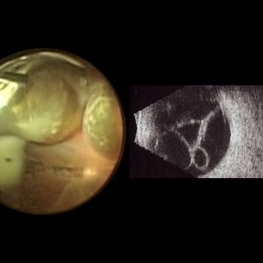
 Eye Finally Got the Ring... But the Retina Was Too Detached to Care
Nov 5 2025 by SHRADDHA RAJ SHRIVASTAVA
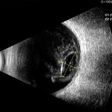
Left Eye B-scan ultrasound of a patient with old retinal detachment shows open funnel shaped hyperechoic membranous echoes, with high amplitude spikes on A-scan and a poor after-movement on dynamic B-scan, suggestive of retinal detachment. We can see a round echogenicity in sub-retinal location, with clear contents within, suggestive of a retinal cyst. This B-scan image is indicative of a long-standing chronic retinal detachment with secondary retinal cyst.
Photographer: Dr. Shraddha Raj Shrivastava
Condition/keywords: B scan ultrasound, chronic retinal detachment, OLD RD, open funnel RD, retinal cyst
-
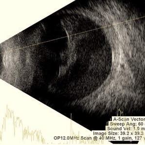
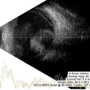
 Closed Funnel RD
Sep 26 2025 by Virginia Gebhart
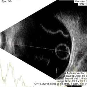
B scan ultrasound of 12 year old male with complete closed funnel RD. Pt endorses several prior blunt traumas which may have caused detachment. If not secondary to trauma, may represent end-stage Coat's disease. FEVR unlikely due to FA findings of fellow eye. Eye is stable since initial visit in 2024, surgical intervention not recommended at this time. Vision NLP
Photographer: Virginia Gebhart, Retina Consultants of Carolina
Imaging device: Keeler Accutome
Condition/keywords: B scan ultrasound, chronic retinal detachment, Closed funnel RD, retinal detachment
-
 B-scan Ultrasound of Choroidal Melanoma with Serous Retinal Detachment
Sep 5 2025 by Kristen Wagner
B-scan ultrasound of a choriodal melanoma with serous retinal detachment.
Photographer: Kristen Wagner, COT Tennessee Retina
Condition/keywords: B scan ultrasound, Choroidal melanoma, serous retinal detachment
-
 Ocular B-scan Ultrasound (Longitudinal Scan M6, gain 100 dB)
Jun 26 2025 by Hector Gabriel Moreno Solano, MD, MHA
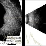
B-scan ultrasound was performed in longitudinal section M6 with a gain of 100 dB. A hyperechoic structure with posterior acoustic shadowing is observed, consistent with lens luxation and condensed vitreous bands adjacent to the lens. The dislocated lens measures approximately 9.54 mm x 4.62 mm. The study was conducted following blunt ocular trauma caused by a golf ball. The remaining vitreous cavity appears anechoic, with no evidence of retinal detachment or other structural abnormalities in this section.
Photographer: Hector Gabriel Moreno Solano, Instituto Mexicano de Oftalmología “IMO I.A.P”
Imaging device: Quantel Medical
Condition/keywords: B scan ultrasound, lens luxation, ocular trauma
-
 Pearl on a String
Apr 28 2025 by rohan jain
Ultrasound of LE of 22 years female showing dislocation of crystalline lens along with retinal detachment
Photographer: Dr. ROHAN JAIN
Condition/keywords: B scan ultrasound, dislocated crystalline lens, Retinal Detachment
-
 Sclerochoroidal Calcification
Apr 24 2025 by Virginia Gebhart
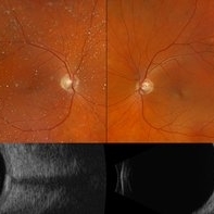
70 year old male referred for amelanotic lesion in the STA OU. Ultrasound shows slightly elevated lesions with hyperreflectivity and posterior shadowing with reduplication artifact consistent with sclerochoroidal calcification. Recommend yearly observation.
Photographer: Virginia Gebhart, Retina Consultants of Carolina
Imaging device: Optos California, Ellex Eye Cubed
Condition/keywords: asteroid hyalosis, B scan ultrasound, sclerochoroidal calcification
-
 USG in Case of Choroidal Hemangioma With Associated Exudative RD
Nov 29 2024 by Anand Temkar
USG of right eye of a 42 year old male, showing us the BRIGHT choroidal hemangioma. This is because they are a mass of relatively large and well-formed blood vessels. Each blood vessel reflects sound waves producing characteristically intense reflections or moderately “MODERATELY HIGH INTERNAL REFLECTIVITY” from within the hemangioma tumor. Moderately high internal reflectivity is an important diagnostic characteristic.
Photographer: Dr.Anand Temkar- Retina Foundation, Ahmedabad
Imaging device: Mirante
Condition/keywords: B scan ultrasound, Choroidal Hemangioma
-
 Macrocysts in Kickboxer
Nov 17 2023 by Bradley T. Smith, MD, FASRS
Intraoperative photo and preoperative b scan of chronic retinal detachment with macrocysts in a kickboxer
Condition/keywords: B scan ultrasound, chronic retinal detachment, ocular trauma, pars plana vitrectomy (PPV), retinal macrocyst
-
 Endophthalmitis
Nov 2 2023 by Anand Temkar
Multiple dot echoes with mild to moderate spikes with free after movements suggestive of vitreous exudates in a case of endophthalmitis.
Photographer: Dr.Anand Temkar- Retina Foundation, Ahmedabad
Condition/keywords: A-scan ultrasound, B scan ultrasound, endophthalmitis
-
 Asteroid Hyalosis
Nov 1 2023 by ANKIT JAIN
B sacn ultrasound image showing hyperechoic echoes with mild to moderate spikes with free after movements on dynamic scan suggestive of asteroid hyalosis.
Photographer: DR ANKIT JAIN
Condition/keywords: asteroid hyalosis, B scan ultrasound
-
 Phthisis Bulbi
Nov 1 2023 by ANKIT JAIN
USG B SCAN image showing multiple dot echos with mild to moderate spikes with free after movements on dynamic scan suggestive of vitreous degeneration, decreased axial length notedand loss of the normal shape of globe suggestive of phthisis bulbi
Photographer: DR ANKIT JAIN
Condition/keywords: B scan ultrasound, phthisis bulbi
-
 Choroidal Melanoma
Nov 1 2023 by ANKIT JAIN
USG B SCAN image showing mass echoes with internal homogeneity with attenuating spikes in a decrescendo pattern likely suggestive of choroidal melanoma.
Photographer: DR ANKIT JAIN
Condition/keywords: B scan ultrasound, CHoroidal melanoma, ultrasound
-
 Retinoblastoma
Nov 1 2023 by ANKIT JAIN
USG B SCAN image showing hyperechogenic mass lesion with moderate spikes with restricted after movements on dynamic scan. In between high spikes noted suggestive of calcification in a case of Retinoblastoma
Photographer: DR ANKIT JAIN
Condition/keywords: B scan ultrasound, retinoblastoma, ultrasound
-
 Choroidal Hemangioma
Nov 1 2023 by ANKIT JAIN
USG B SCAN image showing hyperechogenic mass like lesion with uniform height of spikes through out the mass likely suggestive of choroidal hemangioma
Photographer: DR ANKIT JAIN
Condition/keywords: B scan ultrasound, choroidal hemangioma, ultrasound
-
 Posterior Vitreous Detachment
Nov 1 2023 by ANKIT JAIN
USG B SCAN image showing membranous echoes with low to moderate spikes with free after movements with no attachment to disc suggestive of posterior vitreous detachment.
Photographer: DR ANKIT JAIN
Condition/keywords: B scan ultrasound, posterior vitreous detachment, PVD, ultrasound
-
 POSTERIOR SCLERITIS
Nov 1 2023 by ANKIT JAIN
USG B SACN image showing typical T-sign in axial horizontal view with increased thickening of the sclero-choroidal complex suggestive of posterior scleritis
Photographer: DR ANKIT JAIN
Condition/keywords: B scan ultrasound, posterior scleritis, ULTRASOUND
-
 Intraocular Foreign Body
Oct 31 2023 by Anand Temkar
Echoes with high spikes with restricted aftermovements suggestive od intraocular foreign body
Photographer: Dr.Anand Temkar- Retina Foundation, Ahmedabad
Condition/keywords: A-scan ultrasound, B scan ultrasound, intraocular foreign body
-
 Vitreous degeneration
Oct 31 2023 by Anand Temkar
Few dot echoes with mild to moderate spikes with free aftermovements suggestive of vitreous degeneration
Photographer: Dr.Anand Temkar- Retina Foundation, Ahmedabad
Condition/keywords: A-scan ultrasound, B scan ultrasound
-
 Vitreous Haemorrhage
Oct 31 2023 by Anand Temkar
Multiple dot echoes with mild to moderate spikes with free aftermovements suggestive of vitreous haemorrhage
Photographer: Dr.Anand Temkar- Retina Foundation, Ahmedabad
Condition/keywords: A-scan ultrasound, B scan ultrasound, vitreous blood
-
 Tractional retinal detachment
Oct 29 2023 by Anand Temkar
Membranous echoes with high spikes with restricted after movements suggestive of retinal detachment ( tractional ).
Photographer: Dr.Anand Temkar- Retina Foundation, Ahmedabad
Condition/keywords: A-scan ultrasound, B scan ultrasound, tractional retinal detachment
-
 Subhyaloid Haemorrhage
Oct 29 2023 by Anand Temkar
Multiple dot echoes with mild to moderate spikes with free after movements suggestive of subhyaloid haemorrhage.
Photographer: Dr.Anand Temkar- Retina Foundation, Ahmedabad
Condition/keywords: A-scan ultrasound, B scan ultrasound, subhyaloid blood, SUBHYALOID HEMORRHAGE
-
 Retinal Detachment
Oct 29 2023 by Anand Temkar
Membranous echoes with high spikes with restricted after movements suggestive of retinal detachmment.
Photographer: Dr.Anand Temkar- Retina Foundation, Ahmedabad
Condition/keywords: A-scan ultrasound, B scan ultrasound, open funnel RD
-
 CD
CD
Oct 27 2023 by Anand Temkar
Membranous echoes with moderate to high spikes with restricted after movements suggestive of choroidal detachment.
Photographer: Dr.Anand Temkar- Retina Foundation, Ahmedabad
Condition/keywords: A-scan ultrasound, B scan ultrasound, choroidal detachment
-
 IOL-Drop
Oct 27 2023 by Anand Temkar
High spikes with restricted after movements suggestive of foreign body ( IOL ) in vitreous.
Photographer: Dr.Anand Temkar- Retina Foundation, Ahmedabad
Condition/keywords: A-scan ultrasound, B scan ultrasound, IOL drop
-
 Closed-funnel-RD
Oct 27 2023 by Anand Temkar
Membranous echoes with high spikes with restricted after movements suggestive of retinal detachment ( closed funnel configuration )
Photographer: Dr.Anand Temkar- Retina Foundation, Ahmedabad
Condition/keywords: A-scan ultrasound, B scan ultrasound, Closed funnel RD
 Loading…