-
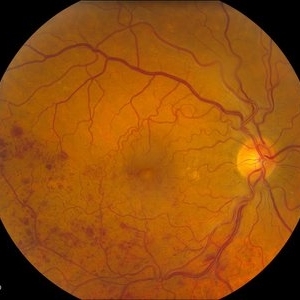
---thumb.JPG/image-square;max$300,300.ImageHandler) Branch Retinal Vein Occulsion
Branch Retinal Vein Occulsion
Jul 10 2013 by Jason S. Calhoun
Patient comes in with complaint of "spot in vision," sort of a cloudy haze in the left eye. Patient's VA was 20/100-1 with no improvement with pinhole in the left eye. Fundus photos show multiple retinal hemorrhages scattered with a branch retinal vein occlusion inferiorily. Patient is also diabetic.
Photographer: Jason S. Calhoun, Department of Ophthalmology, Mayo Clinic Jacksonville, Florida
Condition/keywords: branch retinal vein occlusion (BRVO)
-
---thumb.JPG/image-square;max$300,300.ImageHandler) Branch Retinal Vein Occlusion With Macular Edema
Branch Retinal Vein Occlusion With Macular Edema
Jul 10 2013 by Jason S. Calhoun
Patient comes in with blurry vision in the left eye. Fundus exam shows BRVO with macular edema in the left eye.
Photographer: Jason S. Calhoun, Department of Ophthalmology, Mayo Clinic Jacksonville, Florida
Condition/keywords: branch retinal vein occlusion (BRVO)
-
---thumb.JPG/image-square;max$300,300.ImageHandler) Branch Retinal Vein Occlusion With Macular Edema
Branch Retinal Vein Occlusion With Macular Edema
Jul 10 2013 by Jason S. Calhoun
Patient with blurry vision in the left eye. Fundus exam shows BRVO with macular edema in the left eye.
Photographer: Jason S. Calhoun, Department of Ophthalmology, Mayo Clinic Jacksonville, Florida
Condition/keywords: branch retinal vein occlusion (BRVO)
-
 Branch Retinal Vein Occlusion
Jul 14 2013 by Jason S. Calhoun
Branch retinal vein occlusion in the right eye. Proceeded with Lucentis and will be followed up in 1-month
Photographer: Jason S. Calhoun, Department of Ophthalmology, Mayo Clinic Jacksonville, Florida
Imaging device: TOPCON TRC 50-EX
Condition/keywords: branch retinal vein occlusion (BRVO)
A project from the American Society of Retina Specialists