-
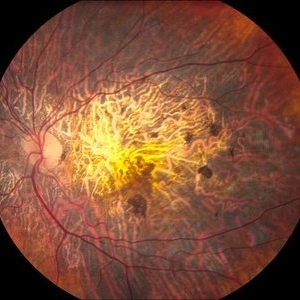 Central Areolar Choroidal Dystrophy
Jun 27 2013 by Jason S. Calhoun
Patient wanted second opinion for atrophic macular degeneration. VA is 20/400, right eye and 20/100, left eye. Patient has very poor vision and is also hearing impaired. Fundiscopic exam reveals very atrophy in the macula. FAF shows a central hole to the choroid with no neovascularization present.
Photographer: Jason S. Calhoun, Mayo Clinic Jacksonville, Florida
Imaging device: TOPCON TRC 50-EX
Condition/keywords: central areolar choroidal dystrophy (CACD)
-
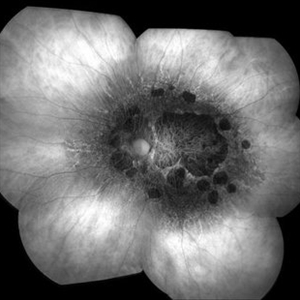 Central Areolar Choroidal Dystrophy
Jun 27 2013 by Jason S. Calhoun
Patient wanted second opinion for atrophic macular degeneration. VA is 20/400, right eye and 20/100, left eye. Patient has very poor vision and is also hearing impaired. Fundiscopic exam reveals very atrophy in the macula. FAF shows a central hole to the choroid with no neovascularization present.
Photographer: Jason S. Calhoun, Mayo Clinic Jacksonville, Florida
Imaging device: TOPCON TRC 50-EX
Condition/keywords: central areolar choroidal dystrophy (CACD)
-
 Central Areolar Choroidal Dystrophy
Jun 27 2013 by Jason S. Calhoun
Patient wanted second opinion for atrophic macular degeneration. VA is 20/400, right eye and 20/100, left eye. Patient has very poor vision and is also hearing impaired. Fundiscopic exam reveals very atrophy in the macula. FAF shows a central hole to the choroid with no neovascularization present.
Photographer: Jason S. Calhoun
Imaging device: TOPCON TRC 50-EX
Condition/keywords: central areolar choroidal dystrophy (CACD)
A project from the American Society of Retina Specialists