-
 Pneumocystis Carinii Choroiditis
Dec 12 2019 by McGill University Health Centre
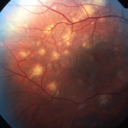
38-year-old HIV positive patient with AIDS. Fundoscopy shows large and small subretinal (choroidal) nodular lesions through out the retina in particular between the arcades.
Photographer: Miguel N. Burnier, McGill University Health Center-McGill University Ocular Pathology & Translational Research Laboratory
Imaging device: Fundoscopy
Condition/keywords: AIDS, choroidal lesions, HIV
-
 Pneumocystis Carinii Choroiditis
Dec 12 2019 by McGill University Health Centre
38-year-old HIV positive patient with AIDS. Fundoscopy showing large and small subretinal (choroidal) nodular lesions throughout the retina in particular between the arcades
Photographer: Miguel N. Burnier, McGill University Health Center-McGill University Ocular Pathology & Translational Research Laboratory
Imaging device: Fundoscopy
Condition/keywords: AIDS, choroidal lesions, HIV, pneumocystis carinii choroiditis
-
 Pneumocystis Carinii Choroiditis
Dec 12 2019 by McGill University Health Centre
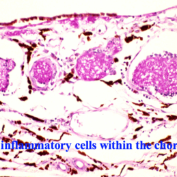
38-year-old HIV positive patient with AIDS. PAS-positive microorganisms at the level of the choriocapillaris and mid-choroid. The histopathological features are consistent with Pneumocystis carinii choroiditis.
Photographer: Miguel N. Burnier, McGill University Health Center-McGill University Ocular Pathology & Translational Research Laboratory
Imaging device: Zeiss
Condition/keywords: choroid, histopathology, pneumocystis carinii choroiditis
-
 Pneumocystis Carinii Choroiditis
Dec 12 2019 by McGill University Health Centre
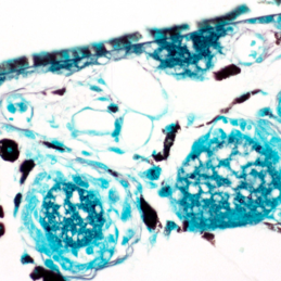
38-year-old HIV positive patient with AIDS. Histopathology showing positive microorganisms (dark- black- round-organisms) by Grocott Methenamine Silver Stain (GMS) in the choroid. The histopathological features are pathognomonic of Pneumocystis carinii choroiditis
Photographer: Miguel N. Burnier, McGill University Health Center-McGill University Ocular Pathology & Translational Research Laboratory
Imaging device: Zeiss
Condition/keywords: choroid, grocott methenamine silver stain, histopathology
A project from the American Society of Retina Specialists