-
 Retinal Capillary Hemangioma
Dec 12 2019 by David L Kilpatrick, MD
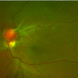
This is a wide-field color fundus photo showing two distinct retinal capillary hemangiomas. A visually significant epiretinal membrane is also present. Work up with gene testing was negative for VHL. The plan is to proceed with PDT of the two separate lesions (half fluence for the peripapillary lesion), followed by cryotherapy / photocoagulation.
Photographer: Retina Consultants of Alabama
Imaging device: Optos
Condition/keywords: retinal capillary hemangioma
-
 Retinal Capillary Hemangioma
Dec 12 2019 by David L Kilpatrick, MD
This is a wide-field color fundus photo showing two distinct retinal capillary hemangiomas. A visually significant epiretinal membrane is also present. Work up with gene testing was negative for VHL. The plan is to proceed with PDT of the two separate lesions (half fluence for the peripapillary lesion), followed by cryotherapy / photocoagulation.
Condition/keywords: retinal capillary hemangioma
-
 Retinal Capillary Hemangioma
Dec 12 2019 by David L Kilpatrick, MD
OCT showing two distinct retinal capillary hemangiomas. A visually significant epiretinal membrane is also present. Work up with gene testing was negative for VHL. The plan is to proceed with PDT of the two separate lesions (half fluence for the peripapillary lesion), followed by cryotherapy / photocoagulation.
Condition/keywords: retinal capillary hemangioma
A project from the American Society of Retina Specialists