-
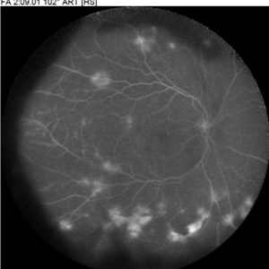 FA 102 PDR
Jul 2 2018 by Lihteh Wu, MD
Ultra wide field fluorescein angiogram of a 59-year-old woman with PDR.
Imaging device: HRA Spectralis Ultra Wide Field
Condition/keywords: proliferative diabetic retinopathy (PDR)
-
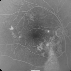
 PDR FA Capillary Non-perfusion
Jul 2 2018 by Lihteh Wu, MD
Fluorescein angiogram of a 59 year old man with PDR. Notice the widespread areas of capillary non-perfusion.
Imaging device: HRA Spectralis
Condition/keywords: proliferative diabetic retinopathy (PDR)
-
---thumb.jpg/image-square;max$300,300.ImageHandler) Coats Disease
Coats Disease
Oct 30 2012 by Lihteh Wu, MD
Fundus photograph of a 29-year-old man with no significant medical or ocular history. Patient complained of progressive loss of vision over the past few months. Notice the lipid exudation over the macula and the hyperplastic RPE.
Condition/keywords: hyperplastic retinal pigment epithelium (RPE), lipid exudation, retinal pigment epithelium
-
---thumb.jpg/image-square;max$300,300.ImageHandler) Coats Disease
Coats Disease
Oct 30 2012 by Lihteh Wu, MD
FA frame showing blocked fluorescence from the massive lipid exudation. There is also hyperfluorescence secondary to vascular leakage and hypofluorescence from the hyperplastic RPE. Superotemporal to the fovea there are areas of telangiectasia.
Condition/keywords: massive lipid exudation, retinal pigment epithelium, retinal telangiectasia
-
---thumb.jpg/image-square;max$300,300.ImageHandler) Coats Disease
Coats Disease
Oct 30 2012 by Lihteh Wu, MD
FA frame showing peripheral telangiectasia and some vascular leakage.
Condition/keywords: peripheral telangiectasia
-
---thumb.jpg/image-square;max$300,300.ImageHandler) Central Retinal Vein Occlusion
Central Retinal Vein Occlusion
Oct 30 2012 by Lihteh Wu, MD
35-year-old hypertensive man with an acute CRVO. Notice the peripapillary cotton wool spots, superficial flame shaped hemorrhages and deeper dot and blot hemorrhages in all 4 quadrants. This is the typical blood and thunder appearance of a CRVO.
Condition/keywords: central retinal vein occlusion (CRVO), cotton wool spots
-
 Melanocytoma
Oct 30 2012 by Lihteh Wu, MD
43-year-old hispanic female found to have on routine examination a melanocytoma of the optic nerve.
Condition/keywords: melanocytoma
-
 Chronic Central Serous Chorioretinopathy
Oct 31 2012 by Lihteh Wu, MD
FA frame showing a hyperfluorescent window defect in a gutter pattern. There is also a hot spot in the nasal macula.
-
 Chronic Central Serous Chorioretinopathy
Oct 31 2012 by Lihteh Wu, MD
FA frame showing a hyperfluorescent window defect in a gutter pattern extending down from the posterior.
Condition/keywords: central serous chorioretinopathy (CSCR)
A project from the American Society of Retina Specialists