-
 Retinal Hypotonia
Oct 15 2025 by Oftalmontt Clínica Láser
Color fundus photograph of a 57-year-old man with streaks on the retinal tapestry suggestive of retinal hypotonia.
Photographer: Ophthalmic Medical Technologist
Imaging device: Canon cx-1
Condition/keywords: retinal hypotonia, Retinography
-
 Retinal Hypotonia
Oct 16 2025 by Oftalmontt Clínica Láser
Vertical B-scan of a 57-year-old male patient showing a retina with multiple folds in all retinal layers, suggestive of retinal hypotonia.
Photographer: Ophthalmic Medical Technologist
Imaging device: RTVue Optovue
Condition/keywords: OCT, retinal hypotonia
-
 Retinal Hypotonia
Oct 16 2025 by Oftalmontt Clínica Láser
Fluorescein angiography of a 57-year-old male patient. The image is presented in arterial time and shows numerous hyperfluorescent folds in the papillomacular sector together with a marked hypofluorescence in the nasal sector.
Photographer: Ophthalmic Medical Technologist
Imaging device: Canon cx-1
Condition/keywords: retinal hypotonia
-
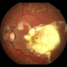 Coats' Disease
Oct 17 2025 by Oftalmontt Clínica Láser
Color retinography of the left eye of a 5-year-old female patient with signs suggestive of unilateral Coats' disease. It presents tortuous telangiectasias associated with a large lipid exudation in the macular and perimacular area together with hyperpigmentation.
Photographer: Ophthalmic Medical Technologist
Imaging device: Canon cx-1
Condition/keywords: Coats' disease
-
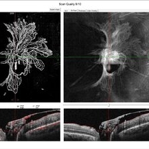 Peripapillary Neovessels
Oct 27 2025 by Oftalmontt Clínica Láser
OCT-A of a 54-year-old male patient with Proliferative Diabetic Retinopathy, observed the large number of neovessels in the optic disc along with fibrovascular proliferation.
Photographer: Ophthalmic Medical Technologist
Imaging device: Avanti XR AngioVue OptoVue
Condition/keywords: diabetic retinopathy
-
 Proliferative Diabetic Retinopathy
Nov 20 2025 by Oftalmontt Clínica Láser
Multimodal examination of a 39-year-old patient presenting with decreased visual acuity in both eyes due to clear proliferative diabetic retinopathy. Presence of neovascularization at the papillary level, microaneurysms in all four quadrants, a vascular loop in the inferior temporal arcade, and an altered ZAF due to low blood perfusion is observed.
Photographer: Ophthalmic Medical Technologist
Imaging device: Canon cx-1 and Avanti XR Optovue
Condition/keywords: proliferative diabetic retinopathy (PDR)
-
 Serous Central Chorioretinopathy With Myelination of Retinal Nerve Fibers
Feb 6 2026 by Oftalmontt Clínica Láser
A 35 year-old patient showed a serous central chorioretinopathy with myelination of retinal nerve fibers in the right eye (temporal fiber raphe can be seen), together with myelination of nerve fibers of the left eye.
Photographer: Ophthalmic Medical Technologist
Imaging device: Canon cx-1
Condition/keywords: central serous chorioretinopathy (CSCR), mielinization
-
 Snowballs Retrocrystalline
Apr 22 2026 by Oftalmontt Clínica Láser
An 8 year-old male patient with pupillary dilation. We can see the presence of "snowballs", a characteristic sign of intermediate uveitis. These are clusters of inflammatory cells, in this case, retrocrystalline snowballs probably attached by fibrin. The retina is applied without major defect, only with the presence of hyperreflective foci in the internal retina.
Photographer: Ophthalmic Medical Technologist
Imaging device: Avanti XR AngioVue OptoVue
Condition/keywords: intermediate uveitis
-
 Long-Standing Toxoplasmosis
Jun 2 2026 by Oftalmontt Clínica Láser
A 12 year-old male patient, accompanied by his guardian, presents for a consultation after being referred by an ophthalmologist due to a macular scar. The guardian reports that the patient has close contact with animals, raising suspicion of toxoplasmosis. The retinography shows the large extent of the long-standing scar, while the OCT reveals a disruption in the organization of the retinal layers, along with the presence of an ERM causing traction and scar tissue
Photographer: Medical technologists in ophthalmology and optometry, OftalMontt
Imaging device: topcon TRC-50DX and Optovue Angiovue OCT
Condition/keywords: ocular toxoplasmosis
-
 Combined Hamartoma of Retina and Retinal Pigment Epithelium
Jun 22 2026 by Oftalmontt Clínica Láser
A 30 year-old female patient underwent OCT, OCTA and retinography, we can see a lesion in the optic nerve with neovascularization, a retinal hamartoma is suspected. It is a rare benign tumor, this lesion is composed of melanocytic tissue (glial and vascular). It is generally thought to be congenital and not hereditary. In the images we can see that the tumor is highly vascularized and that it extends to the foveal sector.
Photographer: Ophthalmic Medical Technologist
Imaging device: Avanti XR AngioVue OptoVue and COMPASS Centervue
Condition/keywords: Combined pigment epithelial and retinal hamartoma
-
 Ciliary Body Cyst
Sep 25 2026 by Oftalmontt Clínica Láser
A middle-aged male patient who underwent LASEK laser refractive surgery attended a postoperative follow-up visit, during which the physician detected iris bulging and subsequently ordered OCT and UBM. The images show iris bulging in the left eye, where UBM visualizes the presence of multiple ciliary body cysts, suggesting that aqueous humor entrapment has occurred.
Photographer: Ophthalmic Medical Technologist
Imaging device: Avanti XR AngioVue OptoVue and UBM ABSolu
Condition/keywords: ciliary body cyst

A project from the American Society of Retina Specialists