-
 Goldmann-Favre Syndrome
Aug 19 2025 by Debarun Sharma
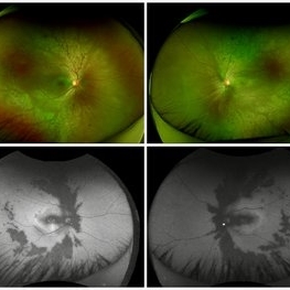
Fundus photograph of a 17 year-old female showing circumferential nummular opacities surrounding the vascular arcades. Fundus autoflourescence shows hypo-autoflourescent circumferential opacities with hyper-autoflourescent ring surrounding macula. Left eye also shows hyper-autoflourescent lesion on the optic nerve head suggestive of astrocytic hamartoma. ERG showed reduced cone response with extinguished rod response. OCT showed schisis of macular area. These features are suggestive of Goldmann-Favre Syndrome.
Photographer: Dr. Debarun Sharma, Sri Sankardeva Nethralaya, Guwahati
Imaging device: Optos
Condition/keywords: Goldmann-Favre Syndrome
A project from the American Society of Retina Specialists