-
 Black Swan - Optic Disc Melanocytoma
Aug 5 2025 by SHRADDHA ASHOK CHANDORKAR, DNB DO FVRS
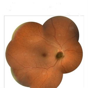
Just like the Black Swan which signifies an event that comes as a surprise, can have a major effect, and is often inappropriately rationalized after the fact with the benefit of hindsight, a 50 yr old presbyopic lady came to OPD with complains of diminution of vision - BCVA being 6/6 N6 in both eyes. Fundus examination revealed a pigmented nodule covering the optic disc .In most cases, fluorescein angiography of a melanocytoma of the optic disk demonstrates hypofluorescence throughout the angiogram. OCT disc showed elevated lesion, OCT macula normal and USG B scan with measurements were done to corroborate the posterior extension and to note increase in size if any on follow ups, perimetry was done to check for any field defects. All tests seemingly within normal limits - Patient was counselled and asked for 6 monthly follow up. Optic Disc Melanocytoma usually unilateral known to be a benign lesion that carries an excellent prognosis, the malignancy of this specific condition is rare 1-2%. The mean age at diagnosis of optic disk melanocytoma is 50 years with a median of 52 and range of 1–91 years. It is possible that melanocytoma is a congenital lesion but may not become clinically apparent until later in life, perhaps due to acquisition of pigment in a previously amelanotic lesion.
Photographer: Dr.Shraddha A. Chandorkar
Imaging device: topcon
Condition/keywords: optic disc melanocytoma
A project from the American Society of Retina Specialists