-
 Choroidal Melanoma
Jul 5 2025 by Gustavo Uriel Fonseca Aguirre
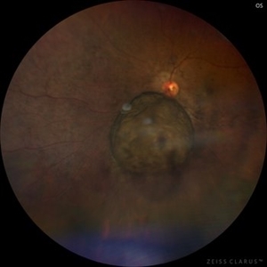
This 50° central fundus photograph reveals a mushroom-shaped choroidal melanoma adjacent to the optic nerve head. The lesion demonstrates characteristic pigmentation with overlying vitreous pigment dispersion (tobacco dust sign).
Photographer: Gustavo U. Fonseca Aguirre, Hospital Conde de Valenciana, Ciudad de México
Condition/keywords: choroidal melanoma
-
 Choroidal Hemangioma (AF)
Jul 5 2025 by Gustavo Uriel Fonseca Aguirre
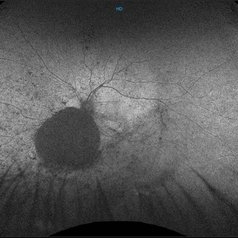
This wide-field fundus autofluorescence image demonstrates a mushroom-shaped choroidal melanoma adjacent to the optic nerve head, exhibiting hypo-autofluorescence (melanin). Vitreous pigment dispersion (tobacco dust sign) is evident, indicating tumor activity.
Photographer: Gustavo U. Fonseca Aguirre, Hospital Conde de Valenciana, Ciudad de México
Condition/keywords: choroidal melanoma
-
 Choroidal Melanoma (USG)
Jul 5 2025 by Gustavo Uriel Fonseca Aguirre
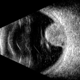
This B-mode transverse ultrasound scan reveals a mushroom-shaped choroidal tumor in the inferior nasal quadrant adjacent to the optic nerve head. The lesion appears solid with homogeneous internal reflectivity and is associated with minimal surrounding subretinal fluid and scleral excavation. It measures 6.54 mm in height × 7.52 mm in base diameter (transverse view) and extends 9.52 mm longitudinally. The vitreous contains abundant punctate opacities consistent with pigment dispersion. The retina and choroid remain attached elsewhere.
Photographer: Gustavo U. Fonseca Aguirre, Hospital Conde de Valenciana, Ciudad de México
Condition/keywords: choroidal melanoma
A project from the American Society of Retina Specialists