-
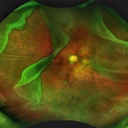 Giant Retinal Tear with Multiple Retinal Breaks
Apr 21 2025 by Hrishikesh Naik, MS
A 28 year old high myope with retinal detachment associated with a supero-temporal giant retinal tear in addition to multiple peripheral horseshoe tears and an additional supero-nasal retinal tear.
Photographer: Hrishikesh Naik
Imaging device: Optos Daytona
Condition/keywords: giant retinal tear, High Myopia, horseshoe tear, retinal break, retinal detachment
-
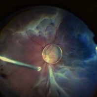 Dislocation of the Crystalline Lens with a Retinal Detachment
Apr 21 2025 by Hrishikesh Naik, MS
An intraoperative screen grab shows a dislocation of the crystalline lens along with an associated rhegmatogenous retinal detachment in a case of Marfan’s syndrome. The case was managed by a combined PPV-SB procedure. A vitrectomy cutter is seen at the left.
Photographer: Hrishikesh Naik
Condition/keywords: intraoperative, lens dislocation, Marfan's syndrome, Retinal Detachment, vitrectomy
-
 Giant Retinal Tear
May 12 2026 by Hrishikesh Naik, MS
A fundus photograph of a 53 year-old with retinal detachment associated with a giant retinal tear.
Imaging device: Zeiss Visucam 224
Condition/keywords: giant retinal tear, retinal detachment
-
 Tractional Retinal Detachment
May 12 2026 by Hrishikesh Naik, MS
Color fundus photograph of a 47 year-old male diagnosed to have advanced diabetic eye disease showing neovascularisation with extensive fibrovascular proliferation with tractional retinal detachment in a table-top configuration. A subretinal napkin-ring band is also seen.
Imaging device: Zeiss Visucam 224
Condition/keywords: proliferative diabetic retinopathy (PDR), Proliferative Diabetic Retinopathy with High-Risk Characteristics, TABLE TOP TRD, tractional retinal detachment, TRD
-
 Advanced Diabetic Eye Disease
Sep 12 2026 by Hrishikesh Naik, MS
A color montage photograph of a 35-year-old man with uncontrolled type 1 diabetes mellitus shows a plethora of features - multiple preretinal, intraretinal and intragel haemorrhages, hard exudates, florid neovascularisation with fibrovascular membranes, posterior pole tractional retinal detachment, ILM folds due to vitreomacular traction and peripheral sclerosed vessels.
Photographer: Maxwell Silva
Imaging device: Zeiss Visucam 524
Condition/keywords: Diabetic fibrovascular membranes, fibrovascular proliferation, neovascular frond, Neovascularisation at the Disc (NVD), preretinal hemorrhage, proliferative diabetic retinopathy (PDR), sclerosed vessels, tractional retinal detachment

A project from the American Society of Retina Specialists