-
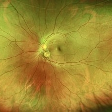 Pseudoduplication of the Optic Disc
Jul 9 2025 by Hrishikesh Naik, MS
A peripapillary colobomatous pseudo-duplication of the optic disc as seen in an asymptomatic 23 year old female with myopia referred for routine retinal periphery screening. Rest retinal exam was normal.
Photographer: Hrishikesh Naik
Imaging device: Optos Daytona
Condition/keywords: Coloboma, Pseudoduplication of optic disc
-
 Double Optic Nerve Shadow
Dec 28 2025 by Hrishikesh Naik, MS
A 10 MHz B-scan ultrasonography image depicting the double optic nerve head shadow sign, the true optic nerve shadow superiorly along with the pseudo optic nerve shadow in the center. The scan reveals a hyperechoic lesion with 100% reflectivity even at a low gain (46 dB) within the posterior outer coats of the eye with posterior acoustic shadowing suggestive of IOFB.
Photographer: Hrishikesh Naik
Imaging device: BioMedix
Condition/keywords: IOFB, Metallic RIOFB
-
 Cytomegalovirus retinitis
May 12 2026 by Hrishikesh Naik, MS
A fundus photograph of a 23 year-old immunocompromised patient presenting with floaters showing a large patch of yellow-white retinal opacification with hemorrhages corresponding to areas of retinal necrosis.
Photographer: Hrishikesh Naik
Imaging device: Zeiss Visucam 524
Condition/keywords: CMV chorioretinitis, CMV retinitis, Cytomegalovirus Retinitis
-
 Valsalva Retinopathy
Jun 1 2026 by Hrishikesh Naik, MS
Montage color fundus photograph of a 22 year-old male following strenuous exercise showing a pre-retinal hemorrhage with a fluid-level sign and an intraretinal hemorrhage superior to the disc, likely suggesting a diagnosis of Valsalva retinopathy.
Imaging device: Zeiss Visucam 224
Condition/keywords: intraretinal hemorrhage, preretinal hemorrhage, subhyaloid hemorrhage, valsalva retinopathy
-
 Frosted Branch Angiitis with Combined Central Retinal Vein Occlusion
Jun 2 2026 by Hrishikesh Naik, MS
Color fundus photograph of a 32 year-old man presenting with sudden diminution of vision in the left eye, showing diffuse retinal periphlebitis in a frosted branch angiitis pattern along with extensive, confluent retinal hemorrhages suggestive of central retinal vein occlusion.
Imaging device: Zeiss Visucam 224
Condition/keywords: central retinal vein occlusion (CRVO), frosted branch angiitis, periphlebitis
-
 Massive Subretinal Hemorrhage Secondary to Polypoidal Choroidal Vasculopathy
Jun 2 2026 by Hrishikesh Naik, MS
Color montage photograph of a 63 year-old male with extensive submacular hemorrhage extending to the near-peripheral retina as a sequelae of polypoidal choroidal vasculopathy (PCV).
Photographer: Hrishikesh Naik
Imaging device: Zeiss Visucam 224
Condition/keywords: polypoidal choroidal vasculopathy (PCV), submacular hemorrhage, subretinal hemorrhage
-
 Healed Cytomegalovirus Retinitis
Sep 11 2026 by Hrishikesh Naik, MS
A fundus photograph of a 23-year-old patient on HA-ART showing a large patch of white retinal opacification corresponding to areas of thinning and gliosis indicative of healed cytomegalovirus retinitis lesion, surrounded by a row of barrage laser marks.
Photographer: Hrishikesh Naik
Imaging device: Zeiss Visucam 524
Condition/keywords: CMV chorioretinitis, CMV retinitis, cytomegalovirus (CMV), Cytomegalovirus Retinitis
-
 Occlusive Vasculitis
Sep 11 2026 by Hrishikesh Naik, MS
Color fundus montage of a 32-year-old man presenting with sudden diminution of vision in the left eye, showing diffuse retinal periphlebitis and occlusive vasculitis in a frosted branch angiitis pattern along with extensive, confluent retinal hemorrhages.
Photographer: Mrinalini Patil
Imaging device: Zeiss Visucam 524
Condition/keywords: central retinal vein occlusion (CRVO), frosted branch angiitis, occlusive vasculitis, periphlebitis

A project from the American Society of Retina Specialists