-
 Choroidal Osteoma
Apr 17 2025 by Gustavo Uriel Fonseca Aguirre
Top (B-mode): The longitudinal scan reveals a hyperechoic, flat, and well-demarcated macular lesion with posterior acoustic shadowing, pathognomonic for choroidal osteoma. Bottom (A-mode): Standardized tracing shows a tall initial spike (100% reflectivity) at the tumor surface with rapid decay to acoustic silence, confirming sound absorption by calcified tissue. This pattern remains unchanged at variable gain settings.
Photographer: Gustavo U. Fonseca Aguirre, Hospital Conde de Valenciana, Ciudad de México
Condition/keywords: choroidal osteoma, macular choroidal osteoma
-
 Choroidal Osteoma
Apr 17 2025 by Gustavo Uriel Fonseca Aguirre
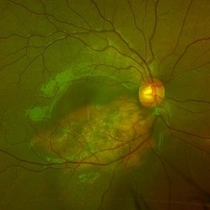
Scanning laser ophthalmoscopy reveals a well-circumscribed, yellowish-white choroidal osteoma overlying the macular region and extending into the inferior temporal vascular arcade. Retinal vessels course normally over the tumor surface, with no evidence of subretinal fluid or hemorrhage. The surrounding retina shows preserved architecture without secondary degenerative changes.
Photographer: Gustavo U. Fonseca Aguirre, Hospital Conde de Valenciana, Ciudad de México
Condition/keywords: choroidal osteoma, macular choroidal osteoma
A project from the American Society of Retina Specialists