-
 Persistent Myelinated Retinal Nerve Fibers
Apr 7 2025 by Juan J. Prados-Carmona
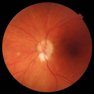
This fundus photograph shows a well-defined, whitish, feathery lesion radiating from the optic disc, consistent with myelinated retinal nerve fibers. The lesion follows the distribution of the retinal nerve fiber layer and appears superficial, partially obscuring the underlying retinal vessels. The optic disc itself is slightly blurred but without signs of true disc edema or hyperemia. The rest of the retina appears unremarkable, with normal vessel caliber and no evidence of hemorrhages, exudates, or signs of hypertensive or diabetic retinopathy. The findings are compatible with a benign congenital anomaly—persistent myelinated retinal nerve fibers.
Photographer: Juan J. Prados-Carmona
Condition/keywords: Persistent myelinated retinal nerve fibers, persistent myelination
A project from the American Society of Retina Specialists