-
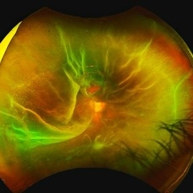 Rhegmatogenous Retinal Detachment
Mar 24 2025 by DR APOORVA JADHAV, MBBS , DNB
This is a color fundus photograph showing rhegmatogenous retinal detachment with posterior pole retinal tear with macula off.
Condition/keywords: retinal detachment of the macula
-
 Proliferative Diabetic Retinopathy
Nov 13 2025 by DR APOORVA JADHAV, MBBS , DNB
Wild Field OCTA of 61 year old male showing us NVD, NVE, CNP areas , macular ischaemia.
Photographer: Dr Apoorva Jadhav
Imaging device: Intalight Dream OCT
Condition/keywords: neovascularization of the disc (NVD), NVE, PDR
-
 Retinal Plumage
Nov 18 2025 by DR APOORVA JADHAV, MBBS , DNB
A 35 year-old male came in with diminished vision in right eye. On multicolor we can appreciate large pigmented choroidal mass lesion with surrounding srf which is reaching fovea. Diagnosed to be Choroidal Melanoma.
Photographer: Dr Apoorva Jadhav
Imaging device: Heidelberg Spectralis
Condition/keywords: choroidal melanoma, srf
-
 OCTA in Branch Retinal Vein Occlusion
Jan 25 2026 by DR APOORVA JADHAV, MBBS , DNB
Wide field OCTA image demonstrating sectoral capillary non perfusion with collaterals in a case of BRVO.
Photographer: Dr Apoorva Jadhav
Imaging device: Intalight Dream Oct
Condition/keywords: CNP areas, ST BRVO
-
 Total Retinal Detachment with Multiple HST
Feb 16 2026 by DR APOORVA JADHAV, MBBS , DNB
Ultra Wide Field fundus photo of a 63 yea- old male showing Total Retinal Detachment with Multiple Horse shoe tears with Inferior PVR changes .
Photographer: Dr Apoorva Jadhav
Imaging device: optos
Condition/keywords: horseshoe tear, Retinal Detachment
-
 Sectoral Silence
Mar 26 2026 by DR APOORVA JADHAV, MBBS , DNB
Wide Field OCTA revealing superotemporal sectoral capillary non perfusion with prominent collateral vascular networks consistent with STBRVO.
Photographer: Dr Apoorva Jadhav
Condition/keywords: cnp, collaterals, starve
-
 Paravenous Pathways
Mar 26 2026 by DR APOORVA JADHAV, MBBS , DNB
Atypical RP variant showing parvenus bone spicule pigmentation with retinochoroidal atrophy diagnosed as Pigmented Paravenous Retinochoroidal Atrophy .
Photographer: Dr Apoorva Jadhav
Condition/keywords: ATYPICAL RP, PPRCA, RP
-
 Neovascular Storm
Mar 26 2026 by DR APOORVA JADHAV, MBBS , DNB
Wide Field OCTA of 54years old male which shows extensive capillary non perfusion areas with multiple tufts of neovascularisation elsewhere exhibiting vascular proliferation at the ischemic margins.Overall features are consistent with PDR .
Photographer: Dr Apoorva Jadhav
Imaging device: Intalight DREAM OCT
Condition/keywords: cnp, nve, pdr
-
 The Silent Intruder: A Metallic Guest in the Retina
Apr 22 2026 by DR APOORVA JADHAV, MBBS , DNB
A 37-year-old male presented with a history of ocular injury from a metallic blade while working with an aluminum fabrication machine. Best-corrected visual acuity was 6/6. Anterior segment examination revealed an inferotemporal full-thickness corneal laceration (3 × 0.5 mm) and a corresponding iris defect. Posterior segment evaluation showed a normal optic disc and macula; however, a highly reflective intraocular foreign body(3x1.5mm) was noted superiorly abutting the retina, with adjacent air bubbles. CT orbit was advised, and a staged surgical plan including primary corneal repair followed by pars plana vitrectomy with foreign body removal was made.
Photographer: Dr Apoorva Jadhav
Condition/keywords: IOFB
-
 Pigmented Paravenous Retinochoroidal Atrophy
Apr 22 2026 by DR APOORVA JADHAV, MBBS , DNB
Atypical RP variant showing parvenous bone spicule pigmentation along the vessels.
Photographer: Dr ApoorvaJadhav
Condition/keywords: RP, RP variant
-
 The Wrinkled Canvas of VKH
Jun 8 2026 by DR APOORVA JADHAV, MBBS , DNB
Wild field fundus photograph of left eye showing multiple serous detachment with choroidal folds and choroidal detachment .Based on clinical and imaging findings , a diagnosis of acute VKH was made . The patient was advised IVMP therapy followed by systemic steroids
Photographer: Dr Apoorva Jadhav
Condition/keywords: choroidal folds, serous detachment, VKH
-
 Radiating Choroidal Folds in Acute VKH
Jun 8 2026 by DR APOORVA JADHAV, MBBS , DNB
Wild field fundus photograph of right eye showing multiple serous detachment with choroidal detachment 360 degrees .Based on clinical and imaging findings , a diagnosis of acute VKH was made . The patient was advised IVMP therapy followed by systemic steroids
Photographer: Dr Apoorva Jadhav
Condition/keywords: serous detachment
-
 A Glowing Core Amid Retinal Tides
Jun 8 2026 by DR APOORVA JADHAV, MBBS , DNB
A 72 year-old female underwent left eye cataract surgery combined with trabeculectomy. Six months later, she presented with decreased vision and was found to have choroidal detachment on examination . BCVA - 6/9 in the affected eye .Fundus evaluation revealed Cdr:0.7 with peripheral 360 degree choroidal elevation .
Photographer: Dr Apoorva Jadhav
Condition/keywords: choroidal detachment

A project from the American Society of Retina Specialists