-
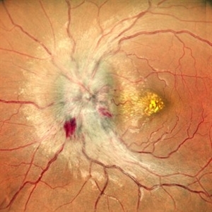
 Pre Retinal Hemorrhage
Jan 11 2025 by rohan jain
Pre retinal hemorrhage
Photographer: Dr. ROHAN JAIN
Condition/keywords: Haemorrhage, hemorrhage, hyaloid membrane
-
 Branches Starved of Flow, Yet Nature Strives to Grow
Apr 1 2025 by rohan jain
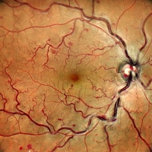
Tufts of NVE's in a case of Branch Retinal Vein Occlusion
Photographer: Dr. ROHAN JAIN
Condition/keywords: branch retinal vein occlusion (BRVO), capillary nonperfusion, non-perfused branch retinal vein occlusion (BRVO), non-perfusion, NVE, OCT Angiography, ST BRVO
-
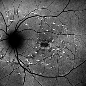
 Craters on the Moon
Apr 21 2025 by rohan jain
Retro image of a 44 year-old woman with Familial Dominant Drusen.
Photographer: Dr. ROHAN JAIN
Condition/keywords: drusen, FAMILIAL DOMINANT DRUSEN, retro mode
-
 Not All Stars Are in the Sky — Some Live in the Eyes of Those Learning to See in New Ways
Apr 21 2025 by rohan jain
Stargardt disease
Photographer: Dr. ROHAN JAIN
Condition/keywords: fleck retinopathy, fundus autofluorescence (FAF), hereditary macular dystrophy
-
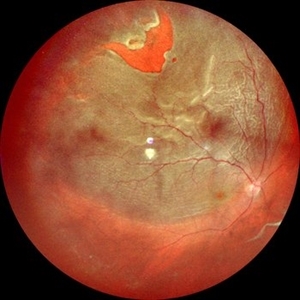 Horse Shoe Tear With Retinal Detachment
Apr 28 2025 by rohan jain
56 year-old male with idiopathic HST and RRD
Photographer: Dr. ROHAN JAIN
Condition/keywords: horseshoe tear, Retinal Detachment, rrd
-
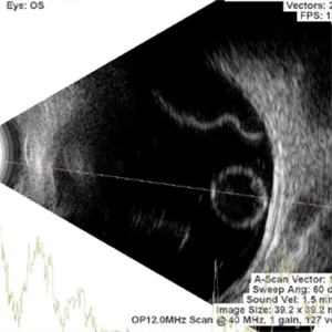 Pearl on a String
Apr 28 2025 by rohan jain
Ultrasound of LE of 22 years female showing dislocation of crystalline lens along with retinal detachment
Photographer: Dr. ROHAN JAIN
Condition/keywords: B scan ultrasound, dislocated crystalline lens, Retinal Detachment
-
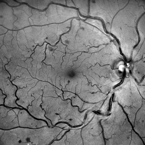 Going with the Flow? Not in CRVO!
Jun 2 2025 by rohan jain
A red free image of central retinal vein occlusion
Photographer: Dr. ROHAN JAIN
Imaging device: mirante
Condition/keywords: central retinal vein occlusion (CRVO)
-
 Blocked and Blurry: A Vein's Midlife Crisis
Jun 2 2025 by rohan jain
A color photograph showing central retinal vein occlusion
Photographer: Dr. ROHAN JAIN
Imaging device: mirante
Condition/keywords: central retinal vein occlusion (CRVO), crvo
-
 When the Macula Decides to Bleed... Artistically (Case of Macular Scar with Subretinal Bleed)
Jun 2 2025 by rohan jain
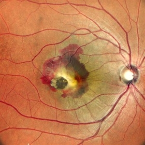
A case of 42 years old male. Color photograph showing macular scar with subretinal bleed.
Photographer: Dr. ROHAN JAIN
Imaging device: mirante
Condition/keywords: CNVM, macular scar, scar, subretinal hemorrhage, subretinal blood
-
 Macular Retinoschisis
Jun 26 2025 by rohan jain
Macular retinoschisis
Photographer: Dr. ROHAN JAIN
Condition/keywords: inferotemporal retinoschisis, juvenile retinoschisis, Macular retinoschisis, RETINOSCHISIS
-
 Macular Retinoschisis
Jun 26 2025 by rohan jain
Macular retinoschisis
Photographer: Dr. ROHAN JAIN
Condition/keywords: juvenile retinoschisis, RETINOSCHISIS
-
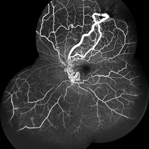
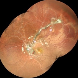
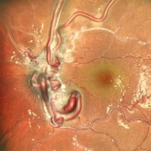
 From Artery to Vein, No Detour: Meet the AV Maverick: Racemose Hemangioma
Jul 1 2025 by rohan jain
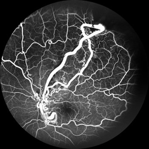
A case of 10 year old girl with defective vision in LE (6/60) who presented us with this condition.
Photographer: Dr. ROHAN JAIN
Imaging device: mirante
Condition/keywords: arteriovenous malformation, FFA in a case of Racemose angioma, racemose hemangioma
-
 From Artery to Vein, No Detour: Meet the AV Maverick: Racemose Hemangioma
Jul 1 2025 by rohan jain
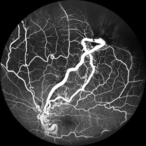
A case of 10 year old girl with defective vision in LE (6/60) who presented us with this condition.
Photographer: Dr. ROHAN JAIN
Imaging device: mirante
Condition/keywords: arteriovenous malformation, FFA in a case of Racemose angioma, racemose hemangioma
-
 From Artery to Vein, No Detour: Meet the AV Maverick: Racemose Hemangioma
Jul 1 2025 by rohan jain
A case of 10 year old girl with defective vision in LE (6/60) who presented us with this condition.
Photographer: Dr. ROHAN JAIN
Imaging device: mirante
Condition/keywords: arteriovenous malformation, FFA in a case of Racemose angioma, racemose hemangioma
-
 Disc Edema with Vasculitis
Jul 15 2025 by rohan jain
Case of disc edema with vasculitis.
Photographer: Dr. ROHAN JAIN
Imaging device: mirante
Condition/keywords: disc edema, idiopathic retinal vasculitis, VASCULITIS
-
 Disc Edema with Vasculitis
Jul 15 2025 by rohan jain
Case of disc edema with vasculitis.
Photographer: Dr. ROHAN JAIN
Imaging device: mirante
Condition/keywords: disc edema, idiopathic retinal vasculitis, VASCULITIS
-
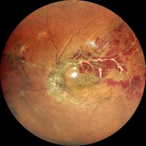
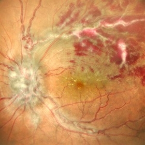
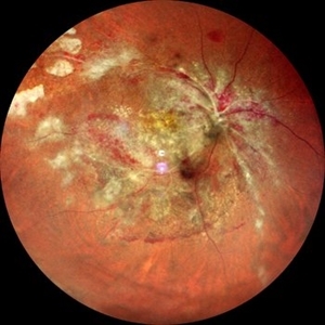
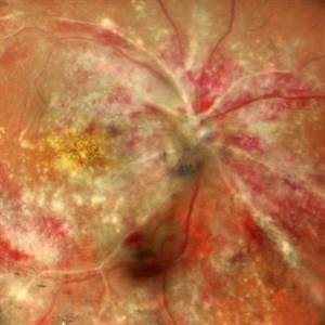 CMV Retinitis: Turning Retina into Abstract Art Since Immunosuppression
Aug 4 2025 by rohan jain
We report a case of 34 years old HIV positive male who presented with Diminution of vision in OD since 1 month. Examination of OD showed hazy media due to vitritis, diffuse yellowish-whitish retinal necrosis and retinal hemorrhages around the disc and attenuated retinal vessels.
Photographer: Dr. ROHAN JAIN
Imaging device: mirante
Condition/keywords: CMV chorioretinitis, CMV retinitis, cytomegalovirus (CMV), Cytomegalovirus Retinitis
-
 CMV Retinitis: Turning Retina into Abstract Art Since Immunosuppression
Aug 4 2025 by rohan jain
We report a case of 34 years old HIV positive male who presented with Diminution of vision in OD since 1 month .Examination of OD showed hazy media due to vitritis, diffuse yellowish-whitish retinal necrosis and retinal hemorrhages around the disc and attenuated retinal vessels.
Photographer: Dr. ROHAN JAIN
Imaging device: mirante
Condition/keywords: CMV chorioretinitis, CMV retinitis, cytomegalovirus (CMV), Cytomegalovirus Retinitis
-
 Bilateral Disc Edema
Sep 11 2025 by rohan jain
bilateral disc edema
Photographer: Dr. ROHAN JAIN
Imaging device: mirante
Condition/keywords: disc edema
-
 Bilateral Disc Edema
Sep 11 2025 by rohan jain
bilateral disc edema
Photographer: Dr. ROHAN JAIN
Imaging device: mirante
Condition/keywords: disc edema
-
 Retained PFCL Over the Optic Disc
Oct 14 2025 by rohan jain
Retained PFCL bubble over the optic disc after retinal detachment surgery.
Photographer: Dr. ROHAN JAIN
Imaging device: mirante
Condition/keywords: near infrared autofluorescence (NIRAF), PFCL
-
 Falciform Retinal Detachment
Nov 22 2025 by rohan jain
Granular fundus with sclerosed retinal vessels with falciform retinal detachment.
Photographer: Dr. ROHAN JAIN
Imaging device: mirante
Condition/keywords: familial exudative vitreoretinopathy (FEVR), persistent fetal vasculature (PFV), persistent hyperplastic primary vitreous (PHPV), Persistent Hyperplastic Primary Vitreous Fibrovascular membrane, ROP
-
 A Galaxy in the Eye—Incidentally Discovered
Jan 27 2026 by rohan jain
A70 year-old male with asteroid hyalosis in left eye.
Photographer: Dr. ROHAN JAIN
Imaging device: mirante
Condition/keywords: ASTEROID, asteroid hyalosis
A project from the American Society of Retina Specialists