-
 Uveal Effusion Syndrome
Jan 7 2025 by Drew Mitchell
Optos Color Montage of Uveal Effusion Syndrome
Photographer: Drew Mitchell, OCT-C
Imaging device: Optos California
Condition/keywords: color photo, montage, OPTOS, uveal effusion
-
 Uveal Effusion Syndrome
Jan 7 2025 by Drew Mitchell
Fundus Autofluorescence Montage of Uveal Effusion Syndrome.
Photographer: Drew Mitchel, OCT-C
Imaging device: Optos California
Condition/keywords: montage, Optos, uveal effusion
-
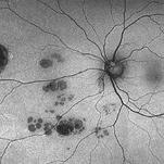 CHRPE and Bear Tracks
Jan 7 2025 by Drew Mitchell
Fundus Autofluorescence of a CHRPE and Bear Tracks.
Photographer: Drew Mitchel, OCT-C
Imaging device: Optos Silverstone
Condition/keywords: bear tracks, CHRPE, congenital hypertrophy of the retinal pigment epithelium (CHRPE)
-
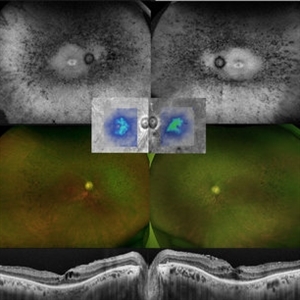 Retinitis Pigmentosa
Feb 18 2025 by Drew Mitchell
FAF, Color, IR, OCT of Mild CME secondary to Retinitis Pigmentosa.
Photographer: Drew Mitchell OCT-C
Imaging device: Optos California
Condition/keywords: cystoid macular edema (CME), Optos, OPTOS CALIFORNIA, retinitis pigmentosa, RP
-
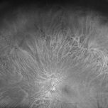 Bear Tracks CHRPE - Red Channel
Jul 29 2025 by Drew Mitchell
Green Free UWF image of extensive bear track patterned CHRPE.
Photographer: Drew Mitchell, OCT-C
Imaging device: Optos California
Condition/keywords: bear tracks, CHRPE, congenital hypertrophy of the retinal pigment epithelium (CHRPE), Green Free, OPTOS CALIFORNIA
-
 Stargardt Disease
Aug 13 2025 by Drew Mitchell
Fundus autofluorescence UWF image taken on a Optos Silverstone of a 44 year old woman with Stargardt Disease.
Photographer: Drew Mitchell OCT-C
Imaging device: Optos Silverstone
Condition/keywords: autofluorescence imaging, Optos, Stargardt Disease
-
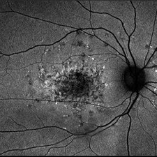
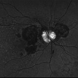
 Optic Disc Drusen
Aug 20 2025 by Drew Mitchell
Optos color photo of a 86 year old woman with neovascular AMD with active CNV and optic disc drusen.
Photographer: Drew Mitchell OCT-C
Imaging device: Optos California
Condition/keywords: color photo, optic disc drusen, OPTOS
-
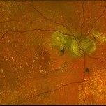
 Optic Disc Drusen
Aug 20 2025 by Drew Mitchell
Fundus Autofluorescence photo of an 86 year old woman with neovascular AMD with active CNV and optic disc drusen.
Photographer: Drew Mitchell OCT-C
Imaging device: Optos California
Condition/keywords: fundus autofluorescence (FAF), neovascular age-related macular degeneration (AMD), optic disc drusen, OPTOS
-
 Coats' Disease
Sep 2 2025 by Drew Mitchell
Optos color photograph of a young boy with Coats disease. Extensive subretinal exudation that is encroaching towards macula. There are peripheral berry aneurysms with localized area of subretinal fluid. Discussed treatment options including laser photocoagulation of aneurysms. Risks benefits and alternatives discussed including possible need for cryo.
Photographer: Drew Mitchell, OCT-C
Imaging device: Optos California
Condition/keywords: Coats' disease
A project from the American Society of Retina Specialists