-
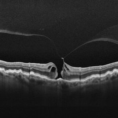
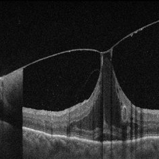 Vitreomacular Adhesion Showcasing a Microaneurysm and a Subhyaloid Hemorrhage
Jan 3 2025 by Drew Mitchell
A vertical OCT 1 line raster scan positioned slightly inferomacula to document the subhyaloid hemorrhage. Hyper reflective Oval indicating Microaneurysm.
Photographer: Drew Mitchell, OCT-C
Imaging device: Zeiss Cirrus 5000
Condition/keywords: diabetic macular edema, microaneurysms, retinal microaneurysms, subhyaloid hemorrhage, subretinal fluid, vitreomacular adhesion, vitreomacular traction (VMT)
-
 Vitreous Hemorrhage (Floater Storm)
Jan 3 2025 by Drew Mitchell
27mm line scan on the Optos Silverstone of a new Vitreous Hemorrhage.
Photographer: Drew Mitchell, OCT-C
Imaging device: Optos Silverstone
Condition/keywords: Optos, swept source, Vitreous hemorrhage
-
 Foveomacular Retinoschisis
Jan 3 2025 by Drew Mitchell
HD 6x6 OCT-Angiography Structural View of the Deep Inner Retina
Photographer: Drew Mitchell, OCT-C
Imaging device: Zeiss Cirrus 6000
Condition/keywords: foveoschisis, maculoschisis
-
 Pattern Dystrophy
Jan 7 2025 by Drew Mitchell
HD 1 line 100x Scan with tracking engaged of Pattern Dystrophy.
Photographer: Drew Mitchel, OCT-C
Imaging device: Zeiss 6000
Condition/keywords: pattern dystrophy, vitelliform lesion
-
 New Subretinal Hemorrhage in AMD
Jan 8 2025 by Drew Mitchell
HD 1 line 100x OCT scan of a New Subretinal Hemorrhage in a established patient with AMD.
Photographer: Drew Mitchell, OCT-C
Imaging device: Zeiss Cirrus 6000
Condition/keywords: age-related macular degeneration (AMD), OCT, subretinal hemorrhage
-
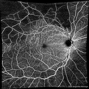 Branch Retinal Vein Occlusion with Macular Edema
Mar 14 2025 by Drew Mitchell
Zeiss Montage Angio 8x8 mm OCT Angiography Superficial Angioplex of a New BRVO in the right eye.
Photographer: Drew Mitchell OCT-C
Imaging device: Zeiss Cirrus 6000
Condition/keywords: branch retinal vein occlusion (BRVO), macular edema, OCT Angiography
-
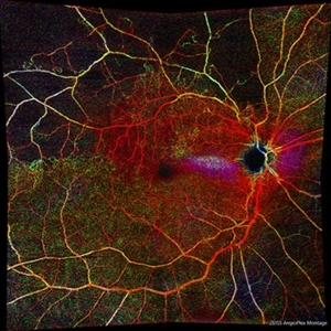 Branch Retinal Vein Occlusion with Macular Edema
Mar 14 2025 by Drew Mitchell
Zeiss Montage Angio 8x8 mm OCT Angiography Retina Depth Encoded Angioplex of a New BRVO in the right eye.
Photographer: Drew Mitchell, OCT-C
Imaging device: Zeiss Cirrus 6000
Condition/keywords: branch retinal vein occlusion (BRVO), macular edema, OCT Angiography
-
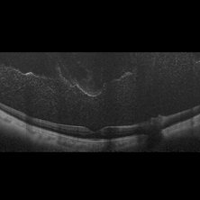
 Myopic Traction Maculopathy
Mar 17 2025 by Drew Mitchell
HD 1 line 100x 9 mm scan of a right eye with MTM at stage 3c. Macular Schisis Detachment.
Photographer: Drew Mitchell OCT-C
Imaging device: Zeiss Cirrus 5000
Condition/keywords: full thickness macular hole, Macular hole, myopic foveoschisis, myopic macular schisis, myopic traction maculopathy, PVD
-
 Retinal Macroaneurysm (RAM)
Mar 19 2025 by Drew Mitchell
3x3 OCT-A of a Retinal Macroaneurysm in the left eye along the IT arcade that has surrounding edema and exudates.
Photographer: Drew Mitchell OCT-C
Imaging device: Zeiss Cirrus 5000
Condition/keywords: OCT Angiography, RAM, retinal macroaneurysm
-
 Retinal Macroaneurysm (RAM)
Mar 19 2025 by Drew Mitchell
3x3 OCT-A of a Retinal Macroaneurysm in the left eye along the IT arcade that has surrounding edema and exudates
Photographer: Drew Mitchell, OCT-C
Imaging device: Zeiss Cirrus 5000
Condition/keywords: CIRRUS 5000 ANGIOPLEX, OCT Angiography, RAM, retinal macroaneurysm
-
 Stage 2 Macular Hole From VMT
Mar 21 2025 by Drew Mitchell
HD 1 line 100x OCT showcasing a full thickness macular hole caused by vitreomacular traction on fovea. Choroidal folds can also be seen on scan.
Photographer: Drew Mitchell OCT-C
Imaging device: Zeiss Cirrus 6000
Condition/keywords: Choroidal Folds, FTMH, macular hole, OCT, PVD
-
 Anastomosis
Jul 29 2025 by Drew Mitchell
3x3 OCT-Angiography Full Depth Color Coded of a left eye with Macular Telangiectasia Type 2
Photographer: Drew Mitchell, OCT-C
Imaging device: Zeiss Cirrus 5000
Condition/keywords: chorioretinal anastomosis, macular telangiectasia type 2, retinochoroidal anastomosis
A project from the American Society of Retina Specialists