-
 Retinal Detachment with Multiple OCT Overlays
Jan 7 2025 by Drew Mitchell
Optos 360* Color photo montage with multiple Zeiss Cirrus OCT scan overlays. Retinal Detachment with multiple breaks and a Epiretinal Membrane.
Photographer: Drew Mitchel, OCT-C
Imaging device: Optos California
Condition/keywords: ERM, macular pucker, montage, Optos, OPTOS CALIFORNIA, RD, Retinal Detachment
-
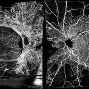 Bilateral Proliferative Diabetic Retinopathy OU
Feb 21 2025 by Drew Mitchell
OCT-Angiography 8x8 Montage OU. PDR with active NVE OD. 37 year old male with no visual complaints. Vision is 20/20 in both eyes.
Photographer: Drew Mitchell OCT-C
Imaging device: Zeiss Cirrus 5000
Condition/keywords: CIRRUS 5000 ANGIOPLEX, Diabetes, NVE, OCT Angiography, proliferative diabetic retinopathy (PDR)
-
 Combined Hamartoma of Retina and Retinal Pigment Epithelium
Aug 13 2025 by Drew Mitchell
Optos color photograph of a 45 year old male with a combined hamartoma of retina and RPE. Epiretinal membrane formation present.
Photographer: Drew Mitchell OCT-C
Imaging device: Optos Silverstone
Condition/keywords: combined hamartoma of retina and RPE, epiretinal membrane formation, ERM, OPTOS, uwf
-
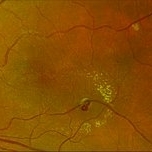 Retinal Macroaneurysm OS (RAM)
Aug 20 2025 by Drew Mitchell
Optos Color photograph of a 79 year old woman with non central macular edema and exudates around RAM inferotemporally.
Photographer: Drew Mitchell OCT-C
Imaging device: Optos Silverstone
Condition/keywords: color photo, exudates, OPTOS, RAM, retinal macroaneurysm
-
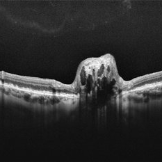 Optic Disc Drusen
Aug 20 2025 by Drew Mitchell
HD 1 line 100x scan through optic disc drusen. ODD are defined as Hyporeflective structures with a full or partial hyperreflective margin.
Photographer: Drew Mitchell OCT-C
Imaging device: Zeiss Cirrus 6000
Condition/keywords: OCT, optic disc drusen
A project from the American Society of Retina Specialists