-
 Oval Pigmented Vitreous Cyst
Nov 27 2024 by Xinyu Zhao
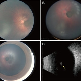
An 8-month-old infant was found to have a brown object in the left vitreous during a fundus screening. A wide-field digital retinal camera (RetCam) revealed a pigmented, non-transparent, freely floating, oval cystic lesion in the vitreous, measuring 2 disc diameters (Figures A-D). The cyst appeared cloudy when focused on the retina (Figure A) but was clearly defined in the vitreous (Figure B). Ultrasound showed a well-defined hyperreflective structure with a hyporeflective lumen (Figure D, indicated by the yellow arrow). A diagnosis of a vitreous pigment cyst, rare in infants, was made. Long-term follow-up is necessary to monitor changes affecting the infant’s vision.
Photographer: Xinyu Zhao, Shenzhen Eye Hospital, Shenzhen, China
Imaging device: RetCam
Condition/keywords: infant, vitreous cyst
A project from the American Society of Retina Specialists