-
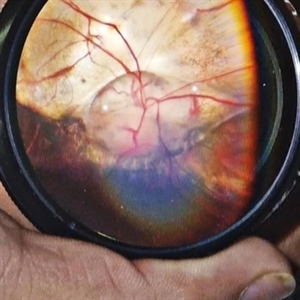
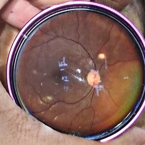 Tuberculoma
Sep 23 2024 by DR Rohit Gupta
Fundus photograph of a 26 year old female suffering from pulmonary tuberculosis, on fundus examination a tuberculoma seen in her left eye.
Photographer: DR Rohit gupta
Imaging device: Samsung S21
Condition/keywords: tubercular choroidal granuloma
-
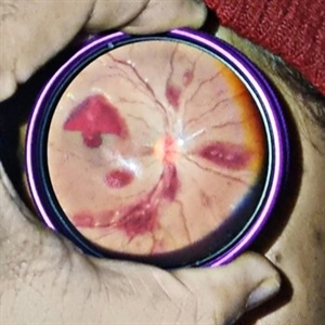 Thrombocytopenia
Sep 24 2024 by DR Rohit Gupta
Fundus photography of a 16 year old female suffering from severe thrombocytopenia. On fundus examination, multiple roth spots and subhyaloid hemorrhage were seen.
Photographer: Dr Rohit gupta
Imaging device: Samsung S21
Condition/keywords: ANEMIC RETINOPATHY, hemorrhage, leukemia, retinal hemorrhage, Roth spots, thrombocytopenia
-
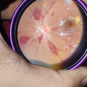 Thrombocytopenia
Sep 24 2024 by DR Rohit Gupta
Fundus photography of a 16 year-old girl suffering from severe thrombocytopenia, showing flame shaped hemorrhage.
Photographer: Dr Rohit gupta
Imaging device: Samsung S21
Condition/keywords: anaemic retinopathy, flame shaped retinal hemorrhage, Haemorrhage, Roth spots, white centered retinal hemorrhage (Roth Spot), white dot syndrome
-
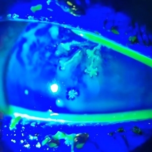 Herpetic Corneal Ulcer
Sep 24 2024 by DR Rohit Gupta
Slit lamp photograph of 32 year old male presented with herpetic corneal ulcer on staining with fluorescein dye under cobalt blue filted dendrits can be seen.
Photographer: Dr Rohit gupta
Imaging device: Samsung S21
Condition/keywords: corneal ulcer, dendritic keratitis, herpes dendrite, Herpes simplex infection, Herpes zoster, staining
-
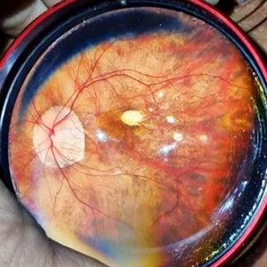
 Fundal Coloboma
Sep 25 2024 by DR Rohit Gupta
Fundus photograph of 16year old female patient with a fundal coloboma in left eye
Photographer: Dr Rohit gupta
Imaging device: Samsung S21
Condition/keywords: chorioretinal coloboma, coloboma of macula, coloboma of optic disc, congenital anomaly
-
 Pathological Myopia
Sep 25 2024 by DR Rohit Gupta
Fundus photograph of a 28 year-old male having high myopia on fundus examination Degenerative changes are seen in retina suggestive of pathological myopia.
Photographer: Dr Rohit gupta
Imaging device: Samsung S21
Condition/keywords: choroidal degeneration, degeneration of optic disc, lacquer cracks, myopia, Myopia macular degeneration CNVM foster fuch spot, pathologic myopia, staphyloma
-
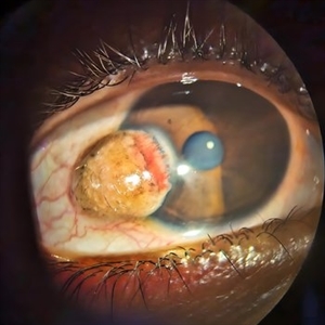 Limbal Dermoid
Sep 25 2024 by DR Rohit Gupta
Slit lamp photograph of a 22 year-old female presenting with a swelling over cornea which on examination appears be to be Limbal dermoid.
Photographer: Dr Rohit gupta
Imaging device: Samsung S21
Condition/keywords: dermoid, limbus, lipodermoid
-
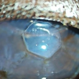 Radial Keratotomy
Sep 28 2024 by DR Rohit Gupta
A slit lamp photograph of 46 year-old female patient operated for high hypermetropia 10 years back . On slit lamp examination hexagonal pattern of radial incisions can be seen.
Photographer: Dr Rohit gupta
Condition/keywords: hypermetropia, hyperopia, Radial keratotomy, refractive surgery
-
 Kayser-Fleischer Ring
Oct 5 2024 by DR Rohit Gupta
Slit lamp photograph of 12 year old male patient suffering from Wilson's disease. This patient presented in opd with extended phalanges, pain in abdomen and disoriented.
Photographer: Dr Rohit gupta
Imaging device: Samsung S21
Condition/keywords: Kayser-Fleischer ring, Kf ring
-
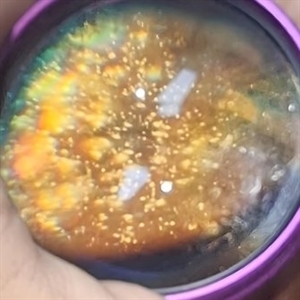 Synchysis Scintillans
Jan 6 2025 by DR Rohit Gupta
60 year old male presented with diminution of vision . On fundus examination multiple crystals deposits seen in vitreous cavity.
Photographer: Dr Rohit gupta
Imaging device: Samsung S21
Condition/keywords: asteroid hyalosis, synchysis scintillans, vitreous cavity
-
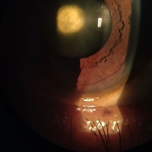
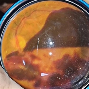 Subhyaloid Hemorrhage
Jan 22 2025 by DR Rohit Gupta
48 year old female presented with right eye diminution of vision, on fundus examination a large hemorrhage was seen in subhyaloid space with multiple retinal hemorrhages. Patients was known case of diabetes with uncontrolled blood sugar level.
Photographer: Dr Rohit gupta
Imaging device: Samsung S21
Condition/keywords: SUB ILM hemorrhage, subhyaloid blood, subhyaloid hemorrhage
-
 PCIOL Opacification
Mar 31 2025 by DR Rohit Gupta
A pseudophakic patient visiting after 6 months of cataract surgery. On slit lamp examination a complete hazy white PCIOL was seen, which is a rare complication after cataract surgery.
Photographer: Dr Rohit gupta
Imaging device: Samsung S21
Condition/keywords: posterior chamber intraocular lens (PCIOL)
-
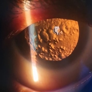
 Posterior Polar Cataract
Apr 10 2025 by DR Rohit Gupta
52 year old male presented with the gradual. Painless, diminuition of vision. On slit lamp examination an onion peel appearance opacification of lens in central part was seen.
Photographer: Dr Rohit gupta
Condition/keywords: posterior capsule opacification, Posterior polar cataract
-
 Myelinated Nerve Fibers
Apr 18 2025 by DR Rohit Gupta
The **myelinated nerve fibers of the optic disc** (also known as **medullated nerve fibers**) are retinal nerve fibers that retain their myelin sheath as they pass through the optic nerve head. Normally, retinal nerve fibers are unmyelinated to allow for light transparency, but in some cases, myelination extends anteriorly into the retina, appearing as a striking white, feathery patch on the optic disc or peripapillary retina. ### **Key Features:** 1. **Appearance:** - Dense, white, striated patches with feathery edges. - Typically located at the superior or inferior pole of the optic disc. - May obscure retinal vessels underneath. 2. **Clinical Significance:** - Usually **benign** and asymptomatic. - **Congenital** (present at birth or early childhood). - Rarely associated with **visual field defects** (e.g., scotomas corresponding to the area of myelination). - Occasionally linked with **high myopia** or **amblyopia** if extensive. 3. **Pathophysiology:** - Failure of oligodendrocytes or Schwann cells to stop myelination at the lamina cribrosa. - Normally, myelination stops at the optic nerve head, but in this condition, it extends into the retina. 4. **Diagnosis:** - **Fundoscopy:** Classic white, feathery appearance. - **Optical Coherence Tomography (OCT):** Shows thickened retinal nerve fiber layer (RNFL). - **Visual Field Testing:** May detect defects if large. 5. **Differential Diagnosis:** - Optic disc edema - Cotton wool spots - Retinoblastoma (rarely, but must be ruled out in children) 6. **Management:** - No treatment required if asymptomatic. - Monitor for amblyopia in children. - Rare cases with significant visual impairment may need further evaluation. ### **Fun Fact:** Myelinated nerve fibers are seen in **~0.5-1%** of the population and are usually an incidental finding.
Photographer: Dr Rohit gupta
Imaging device: Samsung S21
Condition/keywords: Medulated Nerve fibre, Medullated Nerve fibres, myelinated nerve fibers, Myelinated Nerve Fibres, optic disc drusen
-
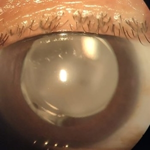
 Posterior Subcapsular Cataract
Apr 18 2025 by DR Rohit Gupta
Slit lamp image on Retroillumination showing posterior subcapsular cataract of a female patient who was on long term corticosteroids inhaler medications.
Photographer: Dr Rohit gupta
Imaging device: Samsung S21
Condition/keywords: posterior capsule opacification, posterior subcapsular cataract, posterior subcapsular changes, posterior subcapsular polar senile cataract, steroids
-
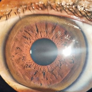
 Iris Hamartomas
Dec 20 2025 by DR Rohit Gupta
Iris photo of a 11 year old female girl presented with multiple iris hamartomas , cafe aeu lait spots , s shaped abaxial proptosis with lid deformity.
Photographer: Dr Rohit gupta
Imaging device: Samsung S21
Condition/keywords: hamartoma, iris nodules, neurofibromatosis
-
 Capillary Hemangioma
Dec 20 2025 by DR Rohit Gupta
A 1 year old male child , presented with his parents , with a red color swelling below eyelid.
Photographer: Dr Rohit gupta
Imaging device: Samsung S21
Condition/keywords: hemangioendothelioma, Hemangioma
A project from the American Society of Retina Specialists