-
 Multimodal Imaging of a Type 3 Retinal Racemose Hemangioma
Sep 8 2024 by Maria Antonia Orrego
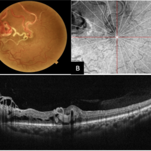
We present the case of a 33 year-old woman with visual loss of her left eye since childhood. Fundus examination revealed a retinal arteriovenous malformation with vessels originating from the optic nerve and extending to the fovea and equator, corresponding to a type 3 retinal racemose hemangioma (A). Infrared reflectance imaging confirmed findings described in funduscopy (B). Spectral domain optical coherence tomography shows dilated vessels in the internal and external retinal layers and adjacent intraretinal fluid (C).
Photographer: Dr. Maria Antonia Orrego V, Universidad CES, Clinica Clofán, Medellín, Colombia
Imaging device: Optovue Solix
Condition/keywords: arteriovenous malformation, multimodal imaging, racemose hemangioma, retinal arteriovenous malformations
A project from the American Society of Retina Specialists