-
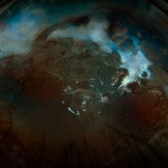 Tractional Detachment of Retina
Aug 21 2024 by Jordyn Beckman
18 year old male with tractional detachment of Retina, chronic macular hole and silicone oil s/p RD repair x2. BCVA CF @2 ft, fellow eye prosthetic.
Photographer: Jordyn Beckman
Imaging device: Optos California
Condition/keywords: Macular hole, preretinal fibrosis, Retinal Detachment, scleral buckle, silicone oil, TRACTION, tractional retinal detachment
-
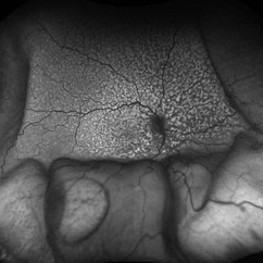 Idiopathic Uveal Effusion Syndrome
Aug 22 2024 by Jordyn Beckman
61 year old male with Idiopathic Uveal Effusion Syndrome with starry night appearance on fluorescein. 3 weeks s/p single external drainage retinotomy and 9 weeks of oral pred with recurrent choroidal effusions. Has since returned to surgery for secondary drainage retinotomy; subretinal fluid remain persistent.
Photographer: Jordyn Beckman
Imaging device: Optos California
Condition/keywords: chorioretinitis, Choroidal, exudative detachment, window defect
-
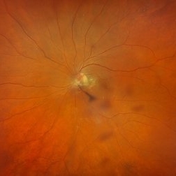 Cloquet Canal
Sep 20 2024 by Jordyn Beckman
79 year old male with wet AMD, no PVD, presents with stable cloquet canal.
Photographer: Jordyn Beckman
Imaging device: Optos California
Condition/keywords: Cloquet Canal, hyaloid membrane, punctum caecum, stilling's canal
-
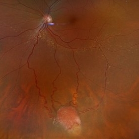 Von Hippel-Lindau Syndrome
Jan 7 2025 by Jordyn Beckman
Fundus photograph of an 37 year old female presents with reddish vascular lesion with feeder vessels for possible Von Hippel-Lindau Syndrome.
Photographer: Jordyn Beckman
Imaging device: California Optos
Condition/keywords: color fundus photograph, feeder vessel, genetic disorder, pre-cryotherapy
-
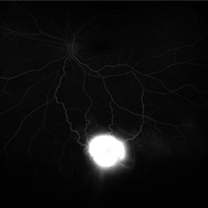 Von Hippel-Lindau Syndrome on FA
Jan 7 2025 by Jordyn Beckman
Fluorescein angiography photograph of an 37 year old female presents with reddish vascular lesion with feeder vessels for possible Von Hippel-Lindau Syndrome.
Photographer: Jordyn Beckman
Imaging device: California Optos
Condition/keywords: feeder vessel, genetic disorder, pre-cryotherapy, Von Hippel-Lindau
-
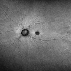 Dome Shaped Macula
Jan 7 2025 by Jordyn Beckman
Autofluorescence photograph of 36 year old woman with Dome Shaped Macula with hypoautofluorescence and hyperautofluorescence centrally.
Photographer: Jordyn Beckman
Imaging device: California Optos
Condition/keywords: autoflorescence, convex protrusion, dome shaped macula, high myopia, hyperautofluorescent centrally, hypoautofluorescence
-
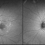 Resolved Multiple Evanescent White Dot Syndrome
Feb 18 2025 by Jordyn Beckman
Autofluorescence fundus photographs of Resolved Multiple Evanescent White Dot Syndrome in a 28 year old female with previous hyperfluorescent punctate spots throughout the posterior pole.
Photographer: Jordyn Beckman, Retina Consultants of Carolina, P.A.
Imaging device: California Optos
Condition/keywords: autofluorescence imaging, grey-white lesions, multiple evanescent white dot syndrome (MEWDS), scattered punctate
-
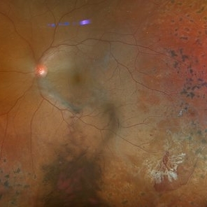 Actively Bleeding NVE
Apr 1 2025 by Jordyn Beckman
47 year old woman presented with actively bleeding NVE temporally on exam with complaints of foggy vision and floaters.
Photographer: Jordyn Beckman, Retina Consultants of Carolina, P.A.
Imaging device: Optos California
Condition/keywords: active bleeding, Elevated retinal neovascularization, vitreous hemorrhage
-
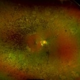 Retinitis Pigmentosa
Apr 1 2025 by Jordyn Beckman
63 year old woman with Retinitis Pigmentosa observed over time with peripheral loss. Over the span of 5 years BCVA changed from 20/25 to 20/50.
Photographer: Jordyn Beckman, Retina Consultants of Carolina, P.A.
Imaging device: Optos California
Condition/keywords: atrophy, bone spicules, retinitis pigmentosa
-
 Amelanotic Lesion with Subretinal Fluid
Mar 9 2026 by Jordyn Beckman
Fundus photograph of a 78 year old man who presented with a central "black spot" in his vision 6 months ago that has been slowly progressing. Lesion is 2.6 mm (H) x 5.7 mm (T) x 4.9 mm (L).
Photographer: Jordyn Beckman, Retina Consultants Of Carolina, P.A.
Condition/keywords: amelanotic, high reflectivity, lesion, secondary macular degeneration, subretinal fluid, subretinal scar
-
 Ciliary Body Melanoma
Mar 9 2026 by Jordyn Beckman
Fundus photograph of a 73 year old woman with a New Ciliary Body Melanoma. UBM showed ciliary body bilobed lesion with low internal reflectivity, most consistent with melanoma. Brachytherapy recommended due to potential for metastatic disease.
Photographer: Jordyn Beckman, Retina Consultants Of Carolina, P.A.
Condition/keywords: bilobed lesion, ciliary body melanoma, low internal reflectivity, plaque placement, possible metastatic risk
-
 Iritis
Jul 28 2026 by Jordyn Beckman
Fundus Fluorescein Angiography of a 80 year old woman with recurrent iritis but was evaluated for birdshot chorioretinopathy. Angiography showed CME, mild late leakage of vessels with no late leakage of nerve with no definite birdshot spots.
Photographer: Jordyn Beckman, Retina Consultants of Carolina
Condition/keywords: cystoid macular edema (CME), Fundus Fluorescein Angiography, iritis, late leakage, normal perfusion, recurrent, RPE mottling

A project from the American Society of Retina Specialists